Silhouette Sign
Cardiac margins are clearly seen because there is contrast between the fluid density of the heart and the adjacent air filled alveoli. Both being of fluid density, you cannot visualize the partition of the right and left ventricle because there is no contrast between them. If the adjacent lung is devoid of air, the clarity of the silhouette will be lost. The silhouette sign is extremely useful in localizing lung lesions.
To utilize the silhouette sign you must know what structures are adjacent to each silhouette.
| Silhouette | Adjacent Lobe/Segment |
| Right diaphragm | RLL/Basal segments |
| Right heart margin | RML/Medial segment |
| Ascending aorta | RUL/Anterior segment |
| Aortic knob | LUL/Posterior segment |
| Left heart margin | Lingula/Inferior segment |
| Descending aorta | LLL/Superior and medial segments |
| Left diaphragm | LLL/Basal segments |
You should know that the pleura encircles the lung and diseases of the pleura can also obliterate silhouettes. The same is true for mediastinal masses.
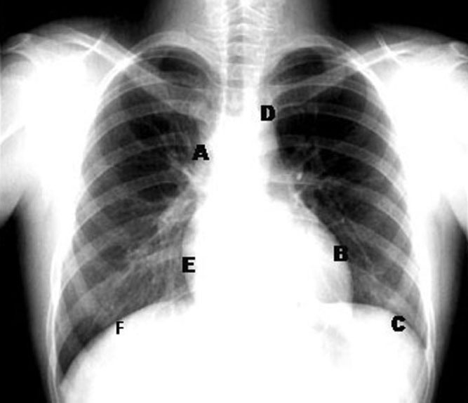 |
A: Ascending aorta B: Left heart margin C: Left diaphragm D: Aortic knob E: Right heart margin F: Right diaphragm
|