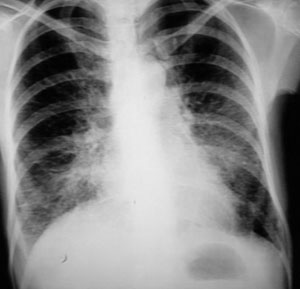
Lymphangitic spread in a patient with cancer breast.
Less than 10% of lung metastases have a lymphangitic pattern.
Pathogenesis
Lymphangitic
metastatic disease in the lung is generally believed to be the result of tumor spread
along the perivascular lymphatic after initial deposition of tumor embolus in a pulmonary
capillary by hematogenous route.
There is evidence that gastric carcinoma is an exception
to this with direct lymphatic extension occurring from the abdomen to chest, across the
diaphragm.
The stomach, lung and breast account for 80% of cases.
The large majority of
patients with unilateral diseases have bronchogenic carcinoma.
Most patients have dyspnea
with or without cough. Initially, symptoms can be mild.
Diagnostic challenge
There is evidence of lung tissue disease on chest radiographs: small linear and
nodular densities, reticular nodular pattern, septal lines.
The appearance is similar to
interstitial changes seen in pulmonary edema, pneumoconiosis, usual interstitial
pneumonitis or sarcoid.
There is frequent pleural effusion on hilar lymphadenopathy.
Some
symptomatic patients have normal radiographs.
Transbronchial lung biopsy or needle aspiration can provide tissue for
diagnosis.
In the absence of suitable chemotherapy, only symptomatic therapy can be
provided.
Most patients become severely dyspneic and expire within a few months.