The lung is filled with air (99% of lung is air), hence, percussion of it gives a resonance. This step helps identify areas of lung devoid of air.
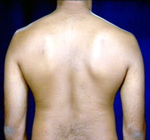
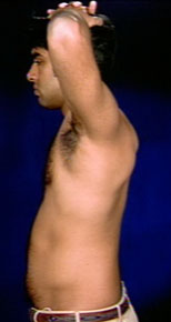
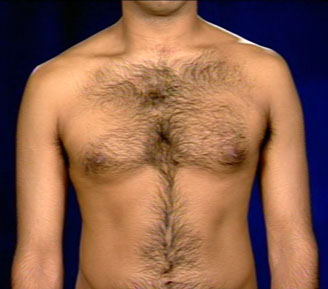
Keep the middle finger firmly over the chest wall along intercostal space and tap chest over distal interphalangeal joint with middle finger of the opposite hand. The movement of tapping should come from the wrist. Tap 2-3 times in a row. Do not leave the percussing finger on , otherwise you will dampen the sound. Listen and feel the resonance. Percuss the chest all around. Stand back, have the patient cross arms to shoulder. This maneuver will wing the scapula and expose the posterior thorax. Percuss starting from top to bottom of thorax on either side. Compare the resonance by percussing the corresponding spaces alternately. Stand on one side and with your flat of hand, tap the chest from top to bottom and from side to side to compare. Then, have the patient keep their hands over head and percuss axilla. Then move to the front and percuss anterior chest , clavicles and supraclavicular space.
Normal
Appreciate the dullness of the left anterior chest due to heart and right lower chest due
to liver. Note the hyper-resonance of the left lower anterior chest due to air filled
stomach. Normally, the rest of the lung fields are resonant.
Abnormal
Decreased or increased resonance is abnormal. Increased resonances can be
noted either due to lung distention as seen in asthma, emphysema, bullous disease or due
to Pneumothorax. Decreased resonance is noted with pleural effusion and
all other lung diseases. Experienced physicians are able to discriminate between dullness
of pleural effusion from a consolidation or a mass lesion of lung. The dullness is flat
and the finger is painful to percussion with pleural effusion.
Example:
Suppose there is dullness half way up the right hemithorax, it certainly rules out the
consideration of pneumothorax. We have to decide whether it is pleural effusion or mass
lesion.