Differential Diagnosis: Crohn's Disease
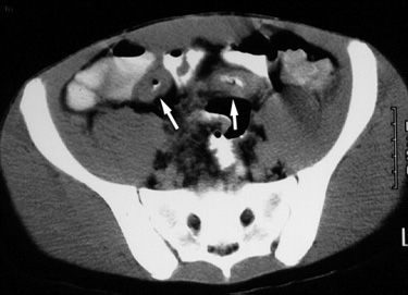
| Crohn's disease mimicking appendicitis. 16 year old male with right lower quadrant pain, rectal pain, and fever for 2 days was evaluated with CT for appendicitis. Several of the CT images are shown below. |
|
Figure 1. CT image through the pelvis after oral contrast administration demonstrates mural thickening of the distal ileum (arrows). There was no evidence of appendiceal abnormality (not shown). |
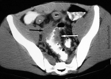 |
Figure 2. CT image, lower in the pelvis than Figure 1, demonstrates marked concentric mural thickening (arrows) involving the rectosigmoid colon. |
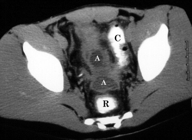 |
Figure 3. CT image at the acetabular level, further caudal than Figure 2, shows two fluid collections (A), consistent with abscesses, anterior to the rectum (R). Note contrast in the adjacent sigmoid colon (C), which improves ability to distinguish between expected (i.e. within bowel) and abnormal fluid collections in the pelvis. |
| The diagnosis of Crohn's disease was made and confirmed by colonoscopic biopsy. |
Related teaching points can also be found in CT Technique and Accuracy
Return to Differential Diagnosis