Differential Diagnosis: Mesenteric Adenitis
Case 1
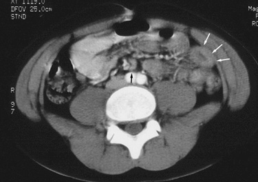
| Mesenteric adenitis mimicking appendicitis. 12 year old male presented to the emergency department with a four day history of colicky right lower quadrant pain. The patient was afebrile, however complete blood count analysis revealed leukocytosis. CT examination, shown below, was requested to evaluate for appendicitis. |
|
Figure 1. CT image just above the iliac crests shows thickened small bowel loops (white arrows) and mildly enlarged mesenteric lymph nodes (black arrow). |
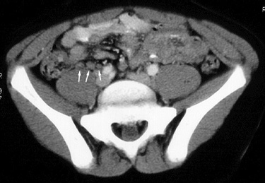 |
Figure 2. CT image slightly lower in the pelvis demonstrates several mildly enlarged right lower quadrant mesenteric lymph nodes (white arrows). The appendix (not shown) was normal in appearance. |
| The patient was diagnosed with non-specific enteritis and mesenteric adenitis. He did well with expectant treatment. |
Return to Differential Diagnosis
Case 2
| Mesenteric adenitis mimicking appendicitis. 18 year old non-pregnant female with right lower quadrant and pelvic pain. On evaluation in the emergency room, the patient was afebrile and had no leukocytosis. Right lower quadrant ultrasound, shown below, was requested to evaluate for appendicitis. |
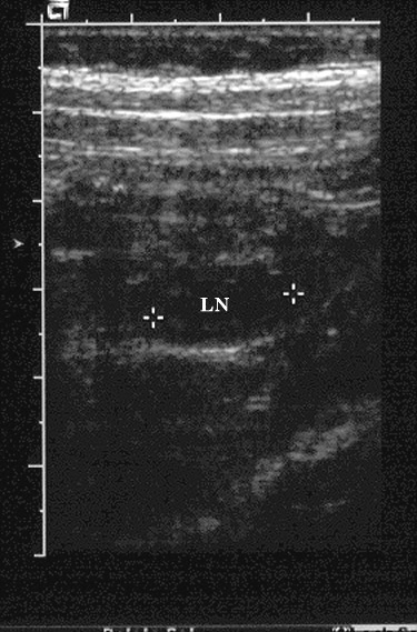 |
Transverse graded compression ultrasound image of the right lower quadrant shows a mildly enlarged mesenteric lymph node (LN). The appendix was not seen; the adnexa (not shown) were normal in appearance. |
| A presumptive diagnosis of mesenteric adenitis was made and the patient did well with no additional treatment. |
Return to Differential Diagnosis