Cerebrovascular Disease (Stroke)

Q1: What are the common causes of stroke?

Cerebrovascular disease can be

Q2: Which imaging procedure should be ordered to evaluate stroke?
Q3: What are the advantages and disadvantages of CT?
Q4: What are the advantages and disadvantages of MRI?
Q5: Does a normal CT or MRI rule out stroke?

Q6: What are the imaging findings of acute infarction?

Acute Infarct:

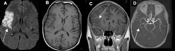
Acute One Day Old Infarction Involving the Right Middle Cerebral Artery (MCA) Territory

A. Diffusion weighted image shows area of infarct as bright signal.
B. T1 image shows no evidence of blood in the area of infarct (blood would appear as white).
C. Post contrast coronal image shows vascular enhancement in the area of infarct.
D. MR angiography shows right middle cerebral artery branches to be narrower in calibre, as compared to left.


Q7: Describe possible subsequent changes on CT following a cortical infarction.

Non contrast CT

MCA distribution infarct seen (hypodense area). Mass effect on the body of the lateral ventricle (arrow).

Three Week Old Subacute Infarct Involving The Right Middle Cerebral Artery (MCA) Territory

A. Diffusion weighted image reveals bright signal involving the cortex. This is from restricted diffusion secondary to acute stroke.
B. Flair image shows bright signal in the posterior parietal cortex with gyral thickening.
C. T1 weighted image shows bright signal in the same area from blood.
D. Post contrast study shows bright signal in the same areas. Enhancement is obscured by the presence of blood.
E. Post contrast study shows bright signal in the same areas. Enhancement is obscured by the presence of blood.

Q8: What are the CT findings of intracerebral hemorrhage (ICH)?

Intracerebral Hemorrhage
Non contrast CT

  • Left parietal hemorrhage with break through into left lateral ventricle.
  • Arrowheads point to hemorrhagic infarct with extension of blood into lateral ventricle (arrow).