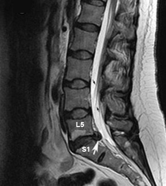
Figure 1: Sagittal T2 wtd. MR image
Disc Herniation
Case 1:
56 year old male with low backache.
Imaging findings: Figure 1
- Extradural compression to the subarachnoid space is seen at L5-S1 level produced by a herniated disc (arrow).
 |
|
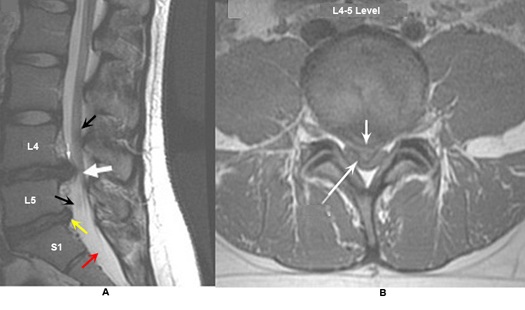
Figure 2: |
|
A. Sagittal T2 wtd. MRI |
B. Axial T1 wtd. MRI |
Disc Herniation
Case 2:
49 year old patient reports with a history of chronic low back pain.
Imaging findings: Figure 2
- Sagittal T2 wtd. MRI of lumbar spine
- The white arrow points to a large herniated disc at L4-L5. CSF appears bright on T2 wtd. MRI (red arrow).
- The yellow arrow points to a smaller disc at L5-S1.
- The black arrows points to nerve roots.
- Axial T1 wtd. MRI of lumbar spine
- The short white arrow points to the herniated disc.
- The long white arrow points to the thecal sac which is compressed ventrally by the herniated disc.
Findings are consistent with a herniated disc.
 |
|
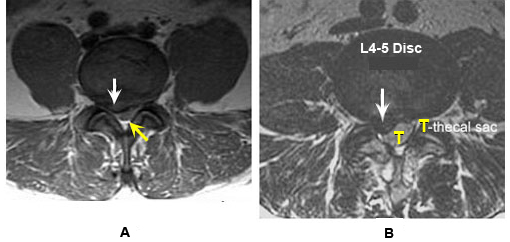
Figure 3: |
|
A. Axial T1 wtd. MRI |
B. Axial T2 wtd. MRI |
Disc Herniation
Case 3:
68 year old male presents with shooting pain along the back of the right leg. He has been having backache for the past six months. His sensation and strength are intact but the right ankle reflex is absent. The pain radiates down into his right buttock and posterior thigh. There is mild weakness of right foot dorsiflexion and toe extension. The patient has a few fasciculations in his right dorsal foot. Pinprick sensation is decreased over his left dorsal foot. There is no Babinski sign. His pain can be reproduced with passive flexion of the hip.
Imaging findings: Figure 3
- Herniated disc (white arrow in A, B)) at L4-L5 level compresses the right side of the thecal sac (yellow arrow in A).
- T for thecal sac (CSF is bright on T2 wtd. image).
Findings are consistent with a herniated disc. Right sided L5 radiculopathy from herniated disc.