 |
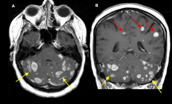
Figure 1 A. Post-contrast axial T1 wtd. MRI |
Multiple Metastatic Tumors to the Brain
Case 1:
40 year old lady with a history of breast carcinoma diagnosed six years ago, presented with headache and ataxia.
Imaging findings: Figure 1
- Shower of numerous metastatic enhancing lesions are seen closely packed together within both cerebellar hemispheres (yellow arrows), and a few lesions also seen within both posterior frontoparietal lobes (red arrows in fig. B).
Final impression:
Multiple metastases to the brain.
Metastatic tumors can be solitary or multiple.
Usually spherical and located at the gray-white matter junction.
Metastatic lesions are better identified by MRI than by CT. Metastatic lesions enhance with gadolinium.
Primary malignant tumors that spread (hematogenous) to the brain include: lung, breast, melanoma, thyroid and renal.
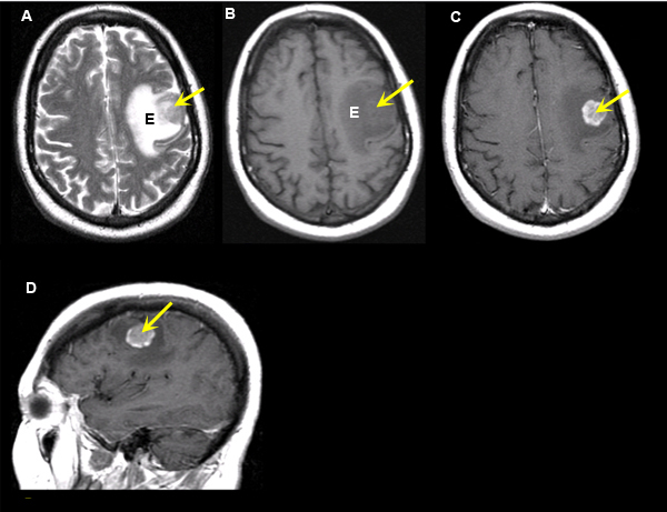 |
Figure 2 A: Axial T2 wtd. MRI |
Solitary metastatic Tumor
Case 2:
67 year old woman status post right mastectomy for breast cancer, presents with severe headache.
Imaging findings: Figure 2
-
Yellow arrows point to the solitary metastatic lesion.
-
Hypointense lesion in the left frontal lobe is seen in pre-contrast axial T1 wtd. MRI (fig. B).
-
Well defined enhancing tumor is seen in post-contrast axial and sagittal T1 wtd. MRI (figs. C, D) surrounded by edema.
-
Edema (E) is seen surrounding the tumor on T1 wtd. image (fig. B), but better appreciated on T2 wtd. image (fig. A) as edema appears bright on this sequence and, thus, readily visible.
Final impression:
Solitary metastatic adenocarcinoma to the brain from breast primary.
Enhancing metastatic tumor is seen with sharply defined margins surrounded by edema in left frontal lobe.
Differential in an elderly patient is Glioblastoma which enhances inhomogenously with large areas of necrosis and irregular margins with finger like projections.
Metastasis is much more common than glioblastoma in adults.