Figure 1
Localized extracellular edema
Case1:
81 year old male with few months of confusion, poor memory, getting lost, misplacing items; left homonymous hemianopsia noted on exam.
Imaging findings:
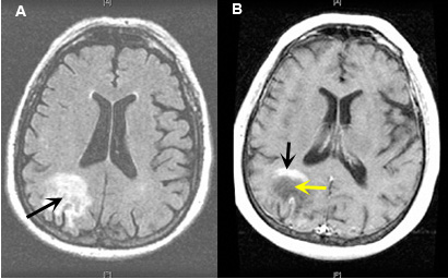
Figure 1: MR images
- FLAIR MR image shows a right parieto-occipital area of high signal from peritumoral edema (black arrow).
- Post-contrast axial MRI: Contrast study helps delineate the lesion causing edema. Edema does not enhance with contrast (yellow arrow). Tumor enhances with contrast (black arrow).
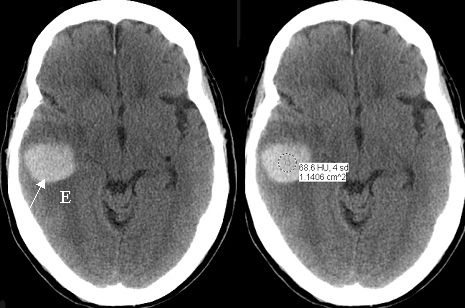
Figure 2
Localized extracellular edema
Case 2:
Image findings:
Figure 2: Pre-contrast axial CT scan
60 year-old patient with melanoma. Hemorrhage is from metastatic tumor bleed.
- Pre-contrast axial CT: Acute intracerebral hematoma within the right temporal lobe (arrow) with surrounding edema (E).
- Pre-contrast axial CT: Acute hematoma is seen by pre-contrast imaging as an area of high attenuation (HU 68.6).
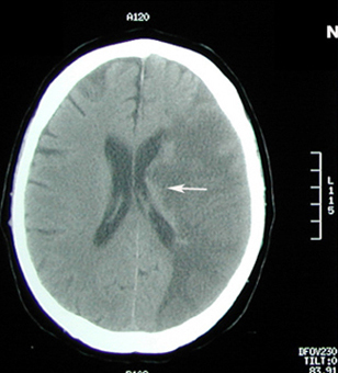
Figure 3
Mass effect producing compression of left lateral ventricles.
Case 3:
Image findings:
Figure 3: Pre-contrast axial CT scan
- MCA distribution infarct with edema is seen (hypodense or low attenuation).
- Mass effect on the body of left lateral ventricle (arrow).
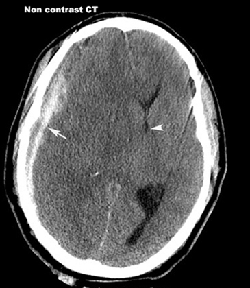
Figure 4
Subdural Hematoma
Case 4:
Image findings:
Figure 4: Pre contrast axial CT scan
- Arrow points to subdural collection of blood (high density).
- Brain edema (hypoattenuation) with shift of right lateral ventricle across the midline (arrowhead) from subfalcine brain herniation.
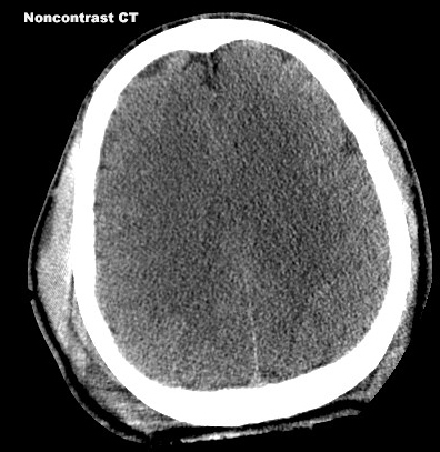
Figure 5
Extracellular edema
Diffuse brain edema
Case 5:
30 year old patient with head trauma.
Image findings:
Figure 5: Pre-contrast axial CT
- Absence of sulcal markings and poor differentiation of white and gray matter from diffuse bilateral cerebral hemispheric edema..
Common causes are head trauma, anoxia, infections, syndrome of inappropriate antidiuretic hormone secretion (SIADH) and high altitude.
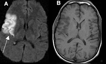
Figure 6
Intracellular cytotoxic edema
Case 6:
Acute one day old infarction involving the right middle cerebral artery (MCA) territory.
Image findings:
Figure 6: MR images
- Diffusion wtd. image shows area of infarct as bright signal (arrow).
- Pre-contrast axial T1 wtd. MRI shows hypodense area in the corresponding site.