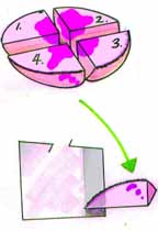
The tissue needs to be mounted so the the entire deep and peripheral margin is all included in one plane, and positioned in the crostat so that the very thin layer that will be shaved off this specimen will contain all the deep and peripheral surgical margin (ie edge of the specimen which was immediately adjacent to the tissue that is still on the patient). So rotate the specimen- (we’ll do #4 first) so that the skin surface portion goes away from you into the screen, and the deepest portion comes up towards you on the screen, and mount in the cryostat in such a way that the part which is staring at you is still staring at you when you look down into the cryostat and its horizontal knife. Click Here to see what this should look like.