Video
of procedure
Takes about 3 minutes
Indications
- Effusion without a secure clinical diagnosis (e.g.,
CHF) or small quantity
- Thoracentesis is a diagnostic procedure done in patients who
have abnormal amounts of fluid accumulation in the pleural space.
- The procedure is usually done at the bedside under local anesthesia.
- The needle is placed through the chest wall into the pleural space and fluid is
then withdrawn into a syringe.
Screening for Thoracentesis
- History of bleeding disorders or use of anticoagulants
- Chest x-ray : Make sure that the fluid is not loculated. This
will have a bearing on the Thoracentesis site.
- Platelet count and PT should be reviewed in patients in whom you
have reason to believe they could be abnormal. It is not necessary to perform them
routinely in other clinical situations.
Premedication
I do not premedicate the patient for a
Thoracentesis.
There is also no need to keep him
NPO.
If you have a high strung patient, you may want
to give him atropine 0.5 mg and 50 mg of Demerol 30 minutes before the procedure.
Selection of Site
The posterior approach is superior.
- The posterior gutter is deep and is
the dependent site where the fluid tends to accumulate in the erect position.
- The interspaces are wider in the back as compared to the front.
- The neurovascular bundle is closer to the inferior margin of the rib
posteriorly. Thus, there is a safer space to enter the chest.
It is scary to see the needle enter the chest. Hence, the anterior approach is
not preferred.
If we select the axillary approach, it is inconvenient to position the patient's
arm for the duration of the procedure. The arm will be in the way.
- It is most comfortable to make the patient straddle a chair.
- He should lean forward on a pillow.
- A nurse can stand in front of the patient and hold the patient's hand.
- We would like to select a site that is dependent and safely away from important
structures. The ideal interspace is the 7th, 8th or 9th space, midway
between the posterior axillary line and midline. This site avoids possible accidental
puncture of the liver, spleen, diaphragm and descending aorta.
- Some experts recommend using the interspace below the upper limit of dullness.
- Of course, in loculated or small effusions, you will select the site most likely
to yield fluid.
Steps of the procedure
Seating
Wing the Scapula
Prepare the Site
Drape
Anaesthetize
Thoracentesis
Return to Menu
options
Complications
The possible complications of Thoracentesis are as follows:
- Pneumothorax: There are two types of pneumothoraxes which can
follow a Thoracentesis.
The first one is secondary to the introduction of air from the outside.
This is benign and does not give rise to any symptoms. It should be left alone.The
second type of Pneumothorax occurs due to an accidental puncture of the lung.
If the patient is asymptomatic, keep him under observation and follow the patient's
progress with a serial chest x-ray. Usually, the puncture in the lung seals and air will
be absorbed spontaneously. If the patient is symptomatic, chest tube drainage may be
necessary.
- Hemothorax: Bleeding is a possibility during a Thoracentesis.
Fortunately, this is rare. Injury to an intercostal artery is fortunately rare since
physicians seem to be aware of their location and avoid it during
Thoracentesis.
- Vaso-Vagal Syncope
- Empyema Empyema is a dreaded complication. Follow strict
surgical aseptic techniques to avoid it.
- Laceration of the Liver or Spleen
- Tumor Seeding Implantation of tumor cells through a
Thoracentesis needle track is an infrequent complication. This occurs with a high degree
of frequency in patients with Mesothelioma and may pose problems. However, with other
tumors, it is of little significance.
- Pain Pain during the procedure is due to poor technique in
the use of a local anesthetic. Occasionally you may encounter a patient who is so high
strung that even touching him may cause pain! Try premedication and reassurance if this
should be the case. Mild pain is to be anticipated for 24 hours after the procedure. If
the patient complains of shoulder pain during the procedure, it indicates that the needle
is piercing the diaphragmatic pleura. The site of the tap is too low.
- Extravasation of Fluid
Subcutaneous Seroma: If the fluid is under tension, extravasation can
occur along the needle track to the subcutaneous tissue. In some patients, this is
massive, disfiguring chest and abdominal wall. Anticipate this complication in massive
effusions, particularly when the fluid spurts out or fills the syringe forcefully during
the Thoracentesis. You may want to release the pressure by evacuating some fluid and
following it up with a firm pressure bandage. Should this occur, reassure the patient.
Usually, it gets reabsorbed in a matter of days.Seneff, et al, carried
out a prospective evaluation of the spectrum and frequency of complications associated
with Thoracentesis. I highly recommend your attention to this article. The following
tables are taken from this reference.
| Major Complications |
| Pneumothorax |
11% |
| Splenic laceration |
0.8% |
| Hemothorax |
0.8% |
| Minor Complications |
| Pain |
22% |
| Cough |
11% |
| Dry tap |
13% |
| Subcutaneous hamatoma |
2% |
| Subcutaneous seroma |
0.8% |
| The reported complications are in a center with trainees! Please be
aware that a Thoracentesis does carry the risk of frequent morbidity. Take every
precaution to minimize its occurrence. It is very safe and pain free in the hands of an
expert. |
Post Thoracentesis Management
Write a procedure note. Be sure to describe the gross appearance of the fluid.
Next you need to consider post procedure orders. The rationale
for post procedure orders are as follows:
- To detect complications
- To evaluate underlying lung
- To distribute specimens
Most physicians consider ordering a Hb and Hct, Chest x-ray, Vital signs
and bed rest.
Following the removal of 50 cc's of fluid for diagnostic purposes, very little
changes occur in the patient's chest x-ray. The underlying lung can be visualized only if
we deliberately evacuated the pleural space.
I do not order any tests routinely following uncomplicated Thoracentesis. I closely monitor the patient's vital signs, CBC and chest x-ray only if one or more of the
following is presented:
- Blood returned in the syringe during the procedure.
- A difficult tap occurred requiring multiple punctures.
- The patient developed symptoms following the tap.
- There is a high risk of bleeding due to a coagulation defect.
- The patient is on a ventilator.
Distribution of specimen
Anticipate all of the required tests and obtain the appropriate
tubes before the actual procedure. Set up priorities for specimen
collection, if something untoward should happen and you are unable to obtain sufficient
amount of fluid, send it for the most important test first.
Studies will be dictated by the clinical diagnosis of the
etiology of the effusion. Please review the lesson on pleural effusion for assistance.
- Tests that should be run (35-50 ml fluid): LDH, protein, WBC
count and differential, glucose, pH; concomitant serum protein, LDH, glucose; arterial pH
if fluid pH is low
- Cultures in appropriate circumstances.
- Special tests appropriate to clinical diagnosis (Lupus,
Mesothelioma etc) <7.30 and acidemia is suspected. Supplement with other reasonably requested analyses cytology, cultures, smears, immunology, amylase, lipids, CEA, etc.
Special Circumstances
Contraindications: none absolute, relative risk > benefit,
bleeding diathesis, small effusion, mechanical ventilation, anticoagulation One must
consider the following special circumstances:
- Loculated Effusion:
The primary concern in loculated effusions is the selection of the Thoracentesis site. The
choice of methods available for site selection are:
- Fluoroscopy
- Ultrasound
- CT
Unless there is Empyema necessitates, it is not a good idea to rely on a
physical examination to select the site of loculation. You will end up puncturing multiple
sites. This is of great pain to the patient. CT is a cumbersome and elaborate test. Ultrasound localization is ideal for this
purpose. It may be done at the bedside. The needle can be placed through the probe and
evacuation can also be ensured in the same sitting.
- Patient on a Ventilator:
There are two considerations for a Thoracentesis when the patient is on a ventilator:
- Risk of Collapsing a Lung:
The fear is whether positive pressure breathing will increase the risk of a puncture to
the lung! My advise is:
- Do not tap small effusions.
- Leave it to an experienced physician.
- Postpone the procedure if the indication is not that urgent.
- Get a post-tap chest film routinely.
- Seating and Positioning:
You will normally be able to position the patient by the side edge of the bed. You can
have the patient rest on an adjustable table. This position will permit you to proceed
with the Thoracentesis in the usual fashion.If you are unable to seat
the patient due to hemodynamic status, mental status or because of tubes and indwelling
lines, the Thoracentesis has to be done in the supine position. Turn the patient on his
side and bring his back to the edge of the bed. You will be able to accomplish the
Thoracentesis in this position.
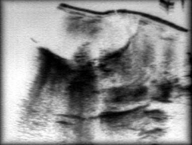
Be aware that the dependent diaphragm moves up. Let me show you a lateral
decubitus chest x-ray to demonstrate the upward movement of the dependent diaphragm.
Select the 5th or 6th interspace to avoid possible injury to the diaphragm. The
selected site should be close to the surface of the bed.
- Patient with a Coagulation Defect:
Postpone the Thoracentesis until the coagulation defect can be corrected. If the defect
cannot be corrected, avoid proceeding with the Thoracentesis. In my opinion, suspected
Empyema will be the only acceptable indication for an emergency Thoracentesis. Leave it to
an experienced physician to perform this procedure. Use a size 21 or 22 needle. Proceed to
attempt with a single stick. Do not give any local anesthetic. Enter the pleural space
with one stroke. Do not try multiple attempts. Closely monitor for a Hemothorax by HGB,
vital signs and a chest x-ray.
Alternate Techniques
There are alternate devices available for
Thoracentesis:
The primary reason for these alternate devices is to circumvent the risk
of a lung puncture by a sharp needle. This occurs during the evacuation process
as the lung expands and meets the needle.
This should not occur with a diagnostic Thoracentesis where only 50 ml of fluid
is removed. If the effusion is small, one should certainly use one of these catheter
devices for Thoracentesis. Of course, one should always use a catheter device to
evacuate the fluid for a therapeutic Thoracentesis.
Utility of test results:
- Exudate vs. transudate:
(1) Fluid/serum protein ratio > 0.5
(2) Fluid/serum LDH ration > 0.6
(3) Fluid LDH > 2/3 upper normal serum LDH; exudates have 1 or more; transudates none
these characteristics
- If LDH only is abnormal - consider malignancy or
Para pneumonic
effusion
- Protein may confuse: e.g., CHF
- <3 g/dl, but might be 3-4 g/dl if patient uses diuretics, or is chronic or recurrent
- WBC: rarely diagnostic alone; > 50,000 in
Para pneumonic
effusion, usually
empyema; > 10,000 very inflammatory
(1) Early, acute, PMN predominant
(2) Later mononuclear - high counts suggest TB, carcinoma, lymphoma, sarcoidosis
(3) Eosinophilia - 10% suggest benign, self- limited; commonly with air or blood in
pleural space; consider: hemothorax, pulmonary infarction, pneumothorax, previous
thoracentesis, parasitic diseases, fungi, drugs, asbestos; rare with TB or malignancy. In
1/3 "idiopathic"
(4) Basophilia - 10%, rare; suggest leukemia
- Mesothelial cells - paucity of cells occurs with chronic diffuse pleural lesions,
e.g., TB, malignancy, empyema rheumatoid effusion, pleurodesis. If > 5%, essentially
rules out TB
- Bloody (> 100,000 cells/mm3): malignancy, trauma, pulmonary embolism,
post-cardiac injury, asbestos pleurisy
- Cytology: yields nearly 90% with malignancy as cause
Transudates
- CHF, cirrhosis, peritoneal dialysis, urinothorax, nephrotic syndrome, atelectasis
Selected Exudates (There are many other causes besides these )
- Para pneumonic - uncomplicated: Clear, LDH<1000, pH> 7.30
- Para pneumonic - complicated: Clear, LDH >1000, pH<
7.30 , Low glucose <40 .
- Empyema- Purulent, LDH>1000, pH < 7.30, Low glucose
<40, Pus cells, demonstrable organisms by stain or culture.
- TB - lymphocytic exudate; pleural biopsy is diagnostic
- Carcinoma - bloody, lymphocytic exudate, cytology or biopsy
positive; if LDH only is abnormal - think cancer
- Esophageal perforation - pH <6.00, high amylase (salivary)
- Rheumatoid pleurisy - turbid, yellow-green, debris- laden fluid;
LDH > 1000, low glucose and Rheumatoid factor ><30, pH 7.00, RF> 1:320
- Lupus - LE cells in effusion (increase if fluid sits up to 24
hours): occasionally low glucose and pH
- Post-cardiac injury syndrome - pleuritic pain, rub, fever 3
weeks after injury; left infiltrates, serosanguineous - no diagnostic labs
- Pulmonary embolism - nothing characteristic; fluid maximal by 72
hours
- Pancreatitis - usually left sided, pleural fluid amylase: serum
amylase > 1.0; amylase may be > 100,000 with pseudocyst
- Asbestos pleural effusion - asymptomatic; bloody exudate,
unilateral , Eosinophilic
- Trapped lung - unilateral; serous, "borderline"
exudate, very low pleural liquid pressure, rapid reaccumulation
- Chylothorax - lymphocytic, milky; chylomicrons in fluid, TG >
110 mg/dl
- Lymphangiomyomatosis - chylothorax in a young women,
interstitial disease, normal lung volumes, repeated pneumothoraces
- Yellow nail syndrome - 40 years old with yellow nails,
lymphedema, respiratory tract involvement, triad not simultaneous; pleurodesis effective
Diagnostic yield
- Almost 75% of thoracentesis yield a specific or presumptive diagnosis; 15-20%
more are useful in management (e.g., rule out empyema)
- Specific diagnoses: malignancy (cells), empyema (pus), tuberculosis pleurisy
(AFB), fungal infection (KOH), lupus pleuritis (LE cells), chylothorax, urinothorax fluid
creatinine/serum creatinine greater than 1), esophageal rupture (high fluid amylase, Ph
about 6.0)