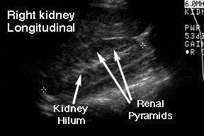
This is a longitudinal view of a right kidney (the bean-shaped structure between the two tiny crosses). Notice that the renal parenchyma is not homogeneous. The cortex (the periphery of the kidney tissue) is grey with some darker circles spaced uniformly around the edge. These darker circles are the renal pyramids. The lighter grey to white area in the center of the kidney is the hilum. This area generates a lot of echoes because there are so many types of tissue in that area: arteries, veins, collecting system, nerves and lymphatics.
Return to G/U Imaging home page.
Return to G/U Development home page.
©David A. Hatch, M.D., 1996