Empyema is defined as accumulation of pus or fluid with demonstrable
bacteria in pleural space.
Clinical Picture
- Patients present with fever, chills, pleuritic chest pain and
cough
- It can be acute , subacute or chronic.
- Leukocytosis with shift to left and Doehle bodies can be noted
on CBC.
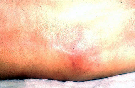
- Besides findings of effusion , clubbing, chest
wall erythema and edema, increased warmth may be noted on physical exam.
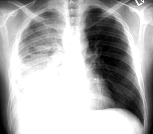
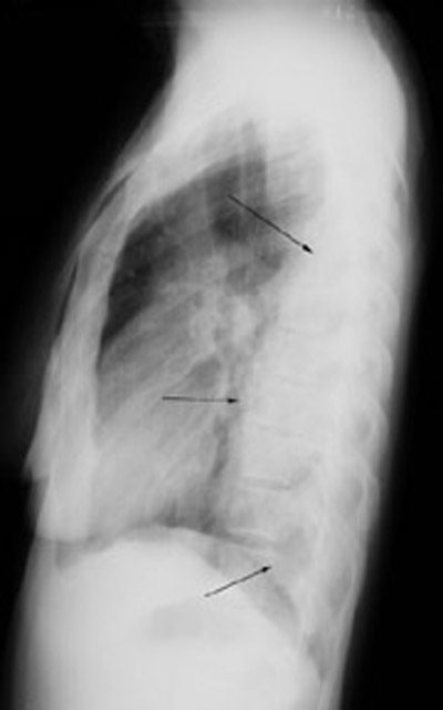
- CXR will
show effusion and cannot be distinguished from other types. Loculated
effusions should raise suspicion for empyema.
- Lack of fever or leukocytosis does not rule out empyema.
Etiology and Pathophysiology
- Empyema most often is due to extension of infection from pneumonia.
Staphylococcal, gram negative and anaerobic infections are common infections presenting in
this mode.
- Anaerobic infections can seed pleura and start as the primary site
of infection without a preceding pneumonitis.
- It could also follow contamination of pleural space from
non-sterile pleural taps.
Diagnosis
- Pleural tap should be done immediately once empyema is a
consideration. If the fluid is grossly
purulent diagnosis is established.
- Gram stain of the pleural fluid and cultures
for aerobes and anaerobes should be obtained.
- If the fluid is not purulent then obtain Ph, glucose and LDH.
This will help categorize parapneumonic effusions as simple and complicated effusions.
- CBC and cultures of sputum and blood are routine.
Treatment
- Empyema should be drained immediately with chest tube
insertion..
- Appropriate Antibiotics should be started immediately, empiric
to start with followed by specific drug based on culture.
- Streptokinase is useful to break up adhesions if there are
loculations.
- Some patients not responding to this regimen may require thoracotomy to lyse
adhesions . This can be accomplished by thoracoscope. Some would require decortication,
if a thick pyemic peel has formed and prevent lung expansion.