The images are not necessarily from this case.
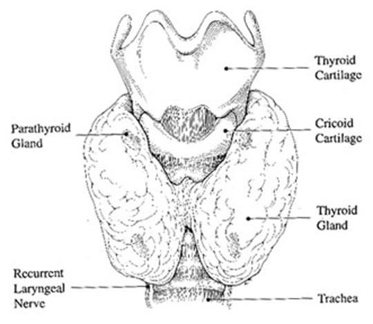
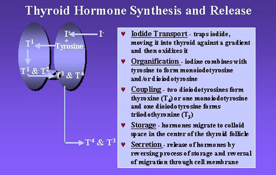
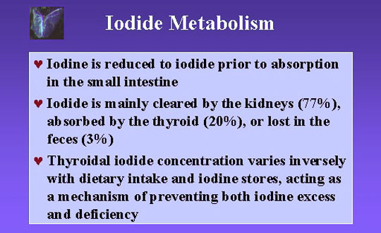
Anatomy, Histology and function
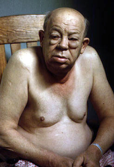
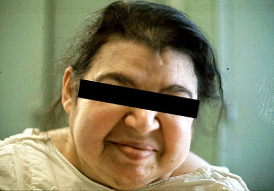
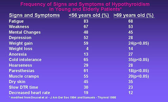
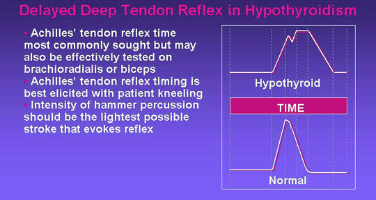
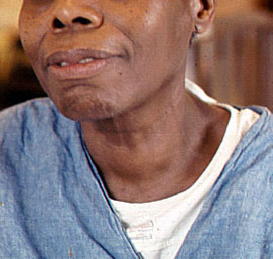
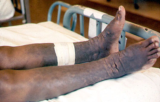
Hypothyroidism
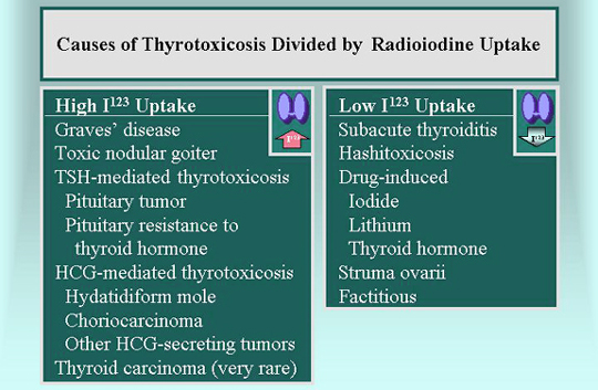
Hyperthyroidism
Pahology
- Operative
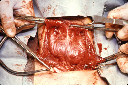
picture: Hyperthyroidism: the photograph depicts the thyroid gland insitu. The thyroid is symmetrically enlarged. Histologic examination revealed hyperplasia modified by pre-operative drugs.
Dr Ralph Leischner
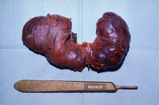
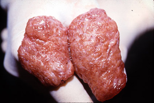
- Hyperthyroidism: the surgical specimen depicts an enlarged thyroid gland removed for hyperthyroidism.
Dr Ralph Leischner
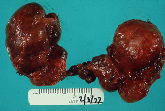
- Hyperthyroidism: the surgical specimen
reveals a bisected thyroid gland. The gland is "beefy" red and firm throughout.
Dr Ralph Leischner
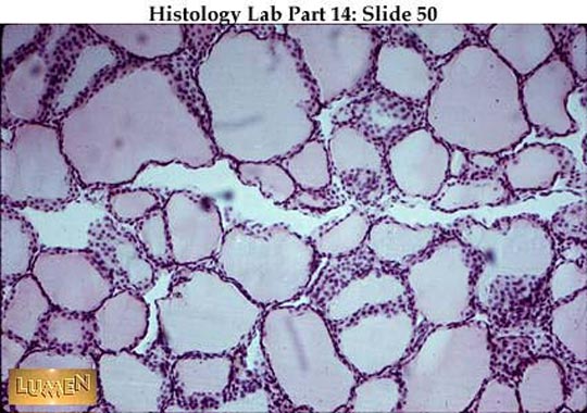
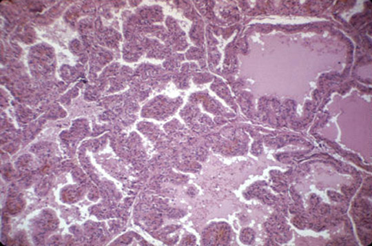
- Hyperthyroidism: microscopic section of the thyroid gland reveals
hyperplasia. Most follicles are lined by hyperplastic epithelium which forms papillary structure. Colloid is decreased is most follicles.
Dr Ralph Leischner
- Multinodular goiter: the surgical specimen reveals an asymmetrically enlarged thyroid gland. The gland is composed of random scattered large and small nodules in both lobes.
Dr Ralph Leischner
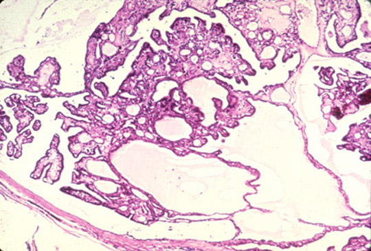
- Multinodular goiter: a microscopic section
of the multinodular goiter reveals colloid distended follicles intermixed with foci of
hyperplasia. Dr Ralph Leischner
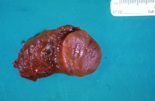
- Thyroid adenoma: the surgical specimen
consists of a lobe of the thyroid gland consisting of adenoma. The adenoma is round, light brown and sharply demarcated from the deep red thyroid tissue.
Dr Ralph Leischner
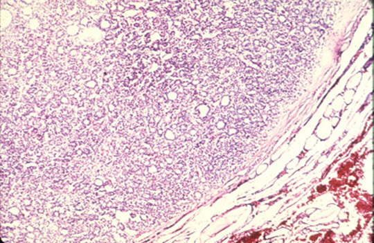
- Thyroid adenoma: a microscopic section
of thyroid gland reveals a follicular adenoma. The neoplasm is composed of small follicles and is surrounded in part by a thin fibrous capsule. The expanding neoplasm compresses the adjacent normal thyroid.
Dr Ralph Leischner
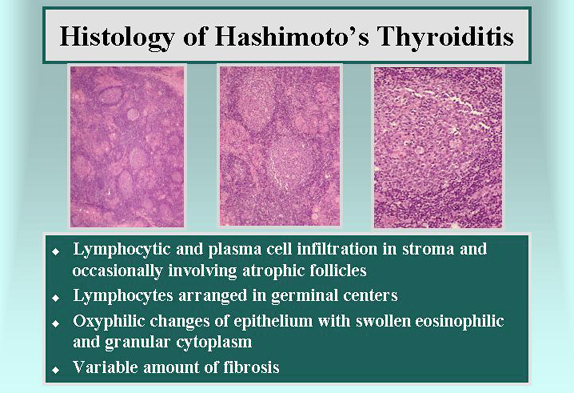
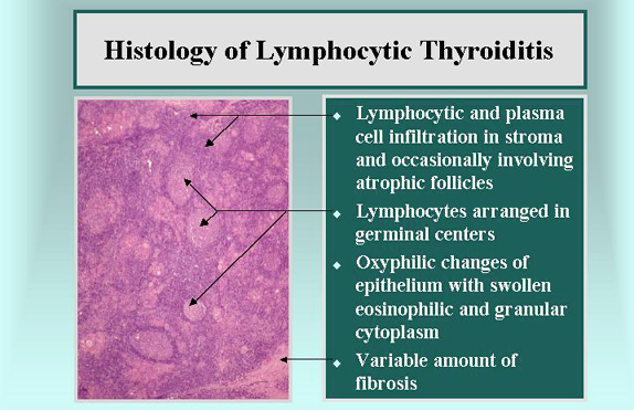
- Hashimotto's
Thyroiditis Dr Donald Gordon
- Viral
thyroiditis Dr Donald Gordon
Send comments to Dr. A.J.
Chandrasekhar M.D.