The images are not necessarily from this case.
Peripheral smear
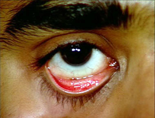
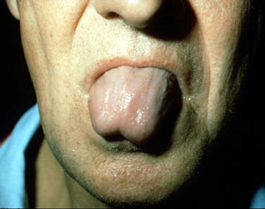
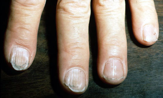
Physical findings
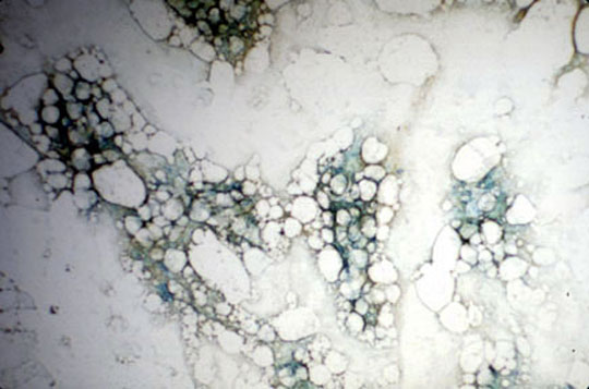
Bone marrow
Reticulocytes
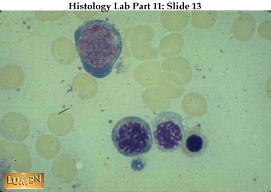
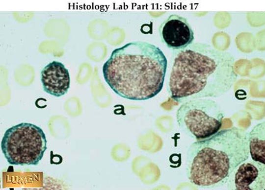
- The cells shown here are all stages in the development of erythrocytes. Generally in the
red blood cell line: (1) the cells become progressively smaller, (2) the cytoplasm
changes from blue to pink, and (3) the nucleus ultimately is lost altogether. Cells shown here
include (in developmental order):
Top cell - proerythroblast
Lower row left = basophilic normoblast or erythroblast. It is still blue, but is smaller; the nucleus is more condensed
middle = polychromatophilic normoblast or erythroblast. Cytoplasm is grayer or muddier; nucleus is even more condensed.
right = orthochromatic (or eosinophilic) normoblast. Cytoplasm is pinker and cell is smaller; nucleus is pyknotic.
Dr John Clancy
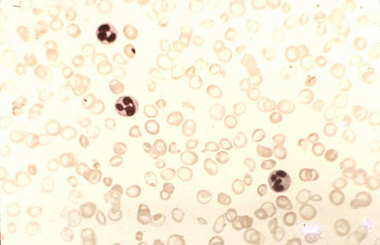
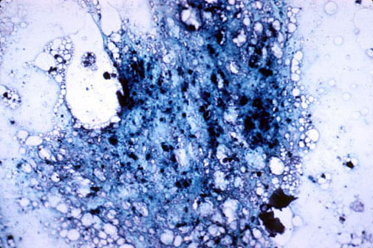
- Reticulocytes with polyribosomal remnants (RNA) staining dark in their cytoplasm. They are slightly larger than the completely mature
erythrocytes and are often found in the peripheral bloodstream at times when blood
cells are being formed unusually rapidly (as during or after certain blood diseases).
Dr John Clancy
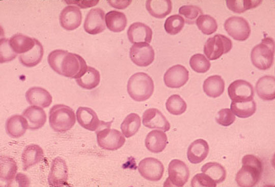
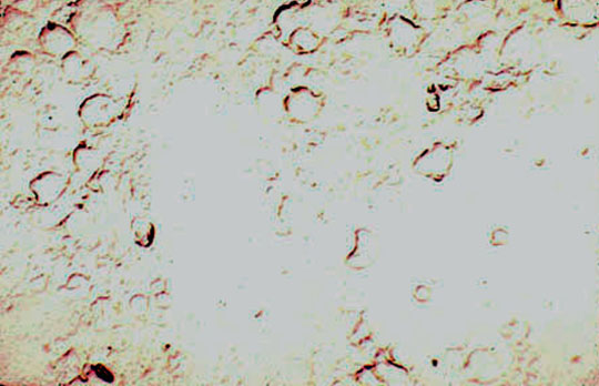
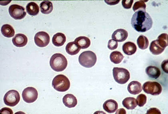
- Blood smear showing polychromasia-a sign of reticulocytosis
Dr Harry Messmore
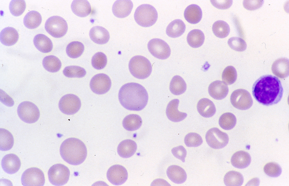
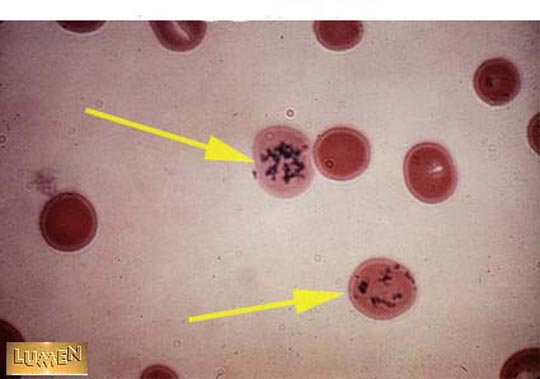
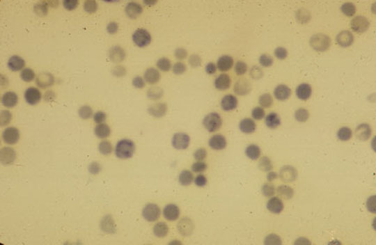
- Reticulocytes in the blood in hemolytic anemia
Dr Harry Messmore
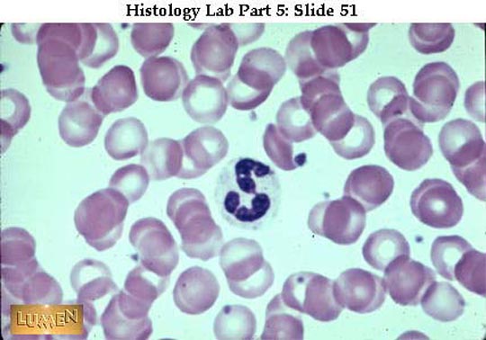
- The young cells in the r.b.c. line all have blue cytoplasm so the you to consider their size in identifying them.
Send comments to Dr. A.J.
Chandrasekhar M.D.