The supportive connective tissue stromar envelopes portal vessels,hepatic artery and bile ducts and enters the liver at its hilus and branches inside the liver to form large septal spaces an smaller portal spaces.
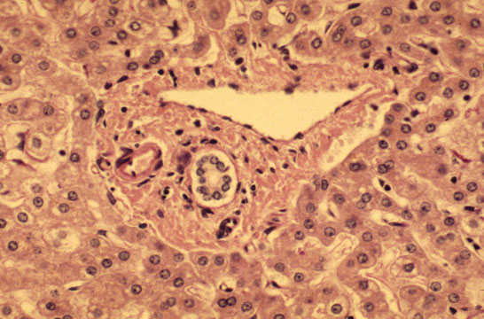 |
PORTAL ARTERIOLE: Located close to the portal bile duct.Small arterioles may penetrate into the lobule.
PORTAL VEIN: Large,thin -walled perforates the limiting plate and connects with sinusoids.Terminates at the equator of the acinus were it forms the terminal portal vessel,not visible in normal conditions.
PORTAL BILE DUCT: Identifiable for its location close to the portal arteriole.
CHOLANGIOLES: Called also bile ductules and ducts of Hering are situated at the periphery of the portal space.
PORTAL LYMPHATICS: Invisible,are around vessels and bile ducts.They drain lymph from the Disse spaces to the hilus of the liver connecting with lymphtics from the glissonian capsule.
INFLAMMATORY CELLS: Presence of lymphocytes and macrophages is normal.Presence of polymorphs, eosinophils and plasma cells is abnormal.
CONNECTIVE TISSUE: Consists of interlacing type I collagen fibers.There is elastic tissue which increases with age.
PERIPORTAL LIMTING PLATE:It is a row of small hepatocytes immediately around the portal connective tissue.
SPACE OF MALL: Space between limiting plate and portal field.It contains villi .
NERVES: Sympathetic from coeliac ganglion and parasympathetic from vagus.They regulate metabolism and vascular and ductal motility.Adrenergic,cholinergic and peptidergic fibers penetrate the lobules where VIP, glucagone,substance P and calcitonin are identified.
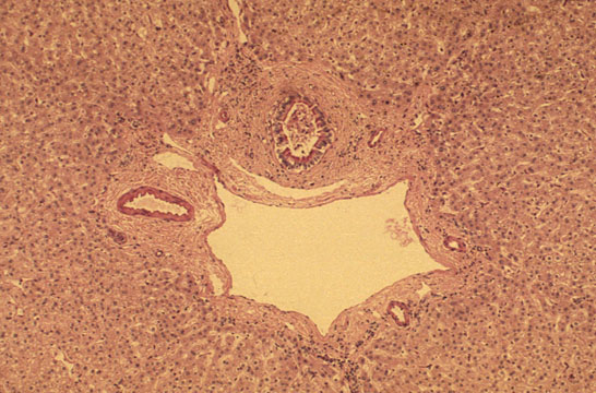 |
Fig 25 - LARGER SEPTAL SPACES: contain larger vessels and bile ducts.Concentric fibrosis around bile ducts in these spaces is normal and should not be interpreted as a pathological change.