|
Pre requisite evaluations |
|||
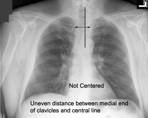
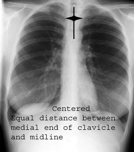
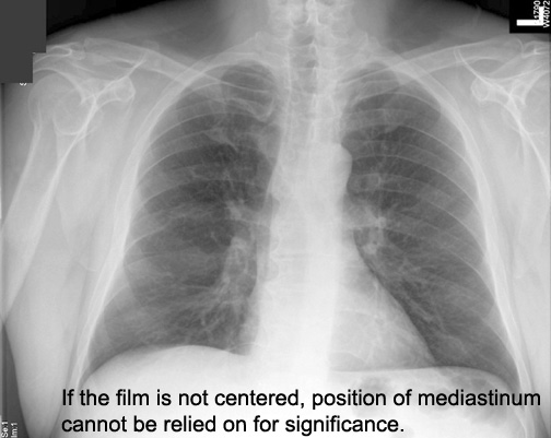
| CXR | Is this film centered? | How do you determine it? Why is that important? | Answer |
| CXR | Is this film centered | Answer | |
| Why is it important to know that the film is centered? | Answer | ||
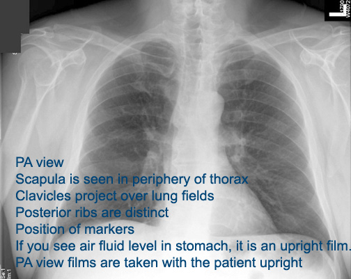
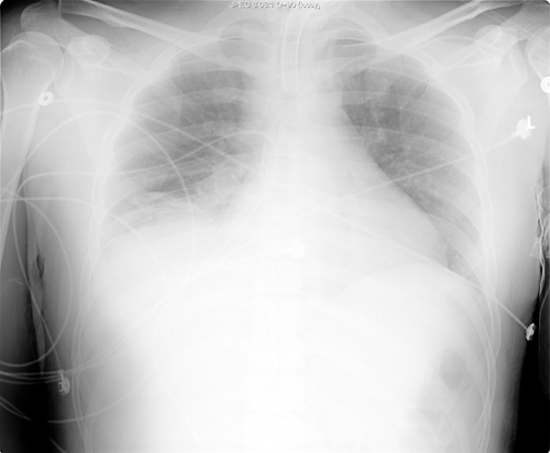
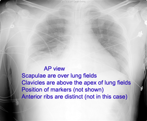
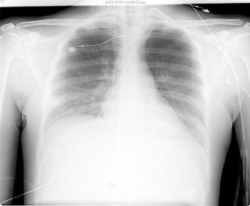
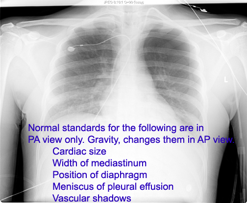
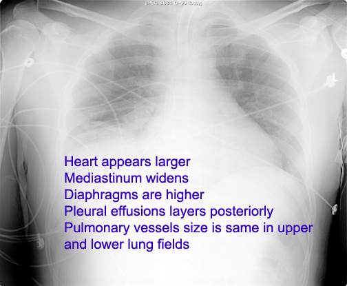
| CXR | Is it PA or AP view? | How can you tell? | Answer |
| CXR | Is it PA or AP view? | How can you tell? | Answer |
| CXR | Is it PA or AP view? | Why is it important to know whether it is PA or AP view? | Answer |
| What are the consideration to be given in interpreting AP view? | Answer | ||
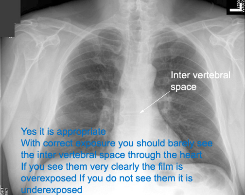
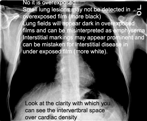
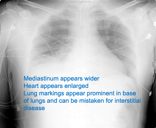
| CXR | Is this film exposed properly? | How do you evaluate exposure? | Answer |
| CXR | Is the exposure appropriate? | Whys is it important to assess exposure? | Answer |
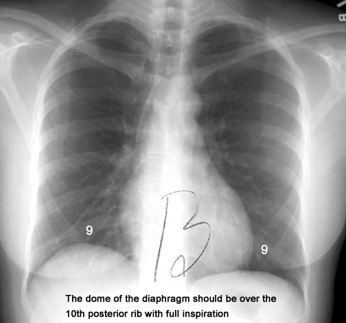
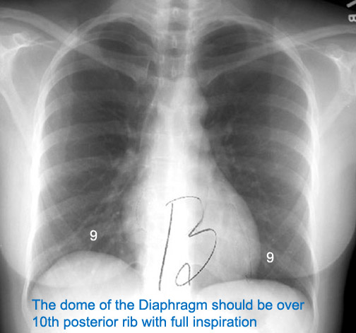
| CXR | Has this patient taken full inspiration? | How do you decide that it is good inspiration film? | Answer |
| What are the problems if the patient has not taken full inspiration? | Answer | ||
|
PA view |
|||
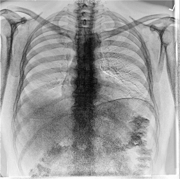
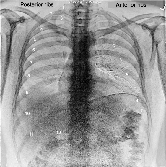
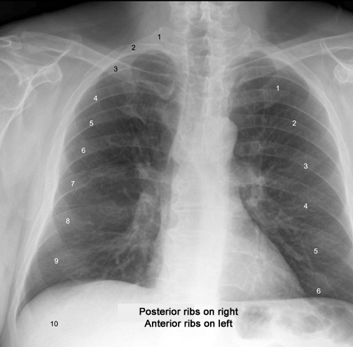
| CXR | Identify the anterior and posterior ribs | Answer | |
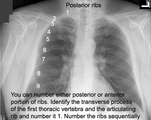
| CXR | How do you number the posterior ribs? | Answer | |
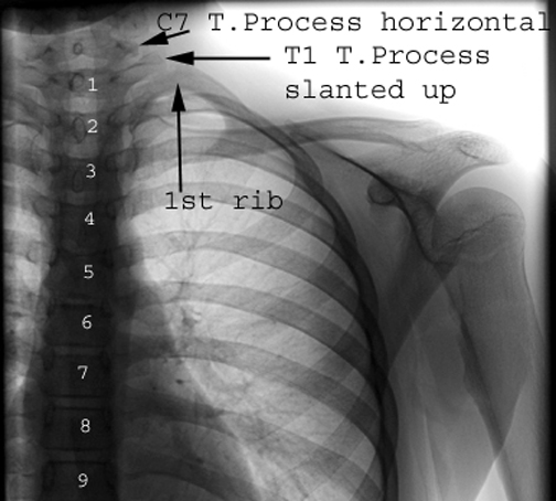
| CXR | How do you identify the first thoracic vertebra? | Answer | |
| CXR | Number the anterior ribs? | Answer Answer | |
| What is the purpose of numbering ribs? | Answer | ||
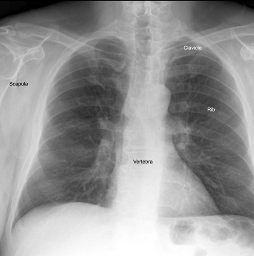
| CXR | Identify Vertebra, scapula, ribs, clavicle | Answer | |
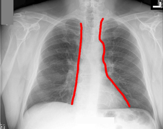
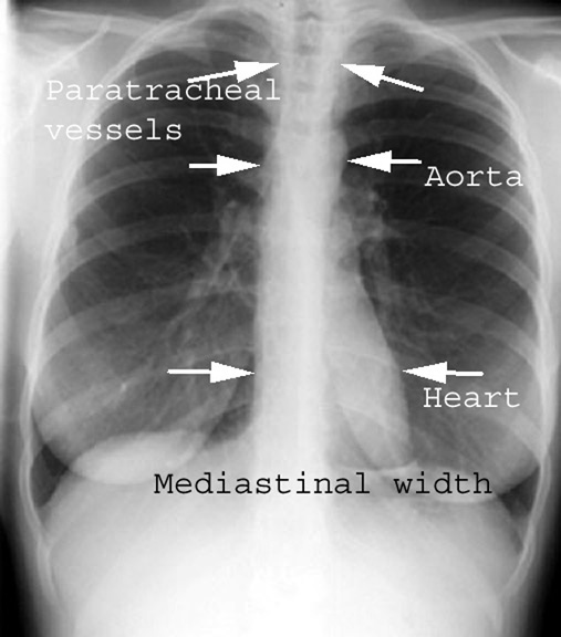
| CXR | Draw the outline of Mediastinum. | Answer | |
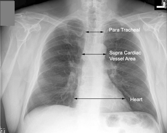
| CXR | What are the Ievels at which you measure mediastinal width? | Answer | |
| CXR | Outline mediastinum | What are the levels at which you measure mediastinal width? | Answer |
| CXR | Outline mediastinum | What is the normal width of mediastinum at tracheal level? | Answer |
| CXR | Outline mediastinum | What is the normal width of mediastinum at supra cardiac vessel area? | Answer |
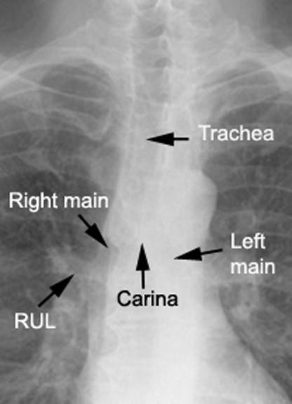
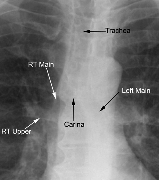
| CXR | Identify Trachea, carina, right and left main stem bronchi. | Why are they visible, while rest of the bronchial tree is not? | Answer |
| What is the carinal angle in this film? | What is the significance of widened carinal angle? | Answer | |
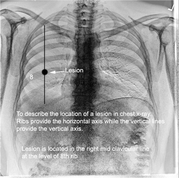
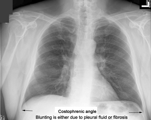
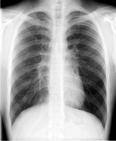
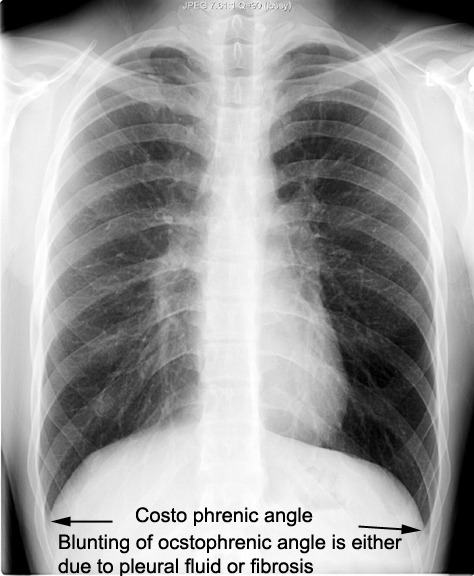
| CXR 8 | Identify costo diaphragmatic (costo phrenic) angles | What does blunting of costo diaphragmatic angle imply? | Answer |
| CXR | Identify costophrenic angles | What does blunting of costophrenic angle imply? | Answer |
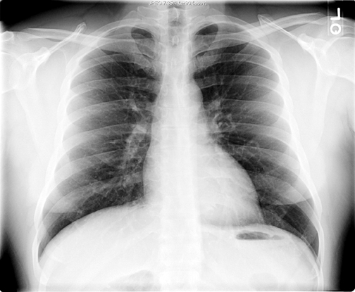
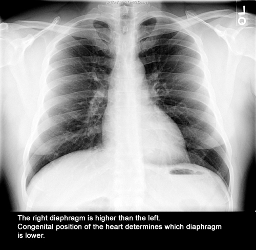
| CXR | Identify diaphragm positions | Which diaphragm is higher and why? | Answer |
| CXR | Identify Trachea, carina, right and left main stem bronchi. | Why are they visible, while rest of the bronchial tree is not? | Answer |
| CXR | Identify right transverse fissure | What is the normal location? | Answer |
| How does location of transverse fissure help you? | Answer | ||
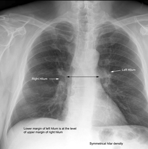
| CXR | Identify left and right hilum. | What is the normal relationship? | Answer |
| How is altered relationship between left and right hilum helpful in diagnostic interpretation? | Answer | ||
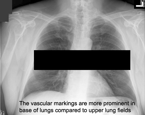
| CXR | Compare and contrast vascular markings in upper vs. lower lung fields in PA view. | Why is there a difference? | Answer |
| List conditions, where vascular markings are prominent in upper lung fields | Answer | ||
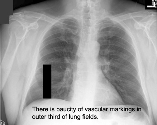
| CXR | What is the difference in vascular markings between outer third and inner two thirds of lungs. | Answer | |
| What is the significance of increased markings in outer third of lung fields? | Answer | ||
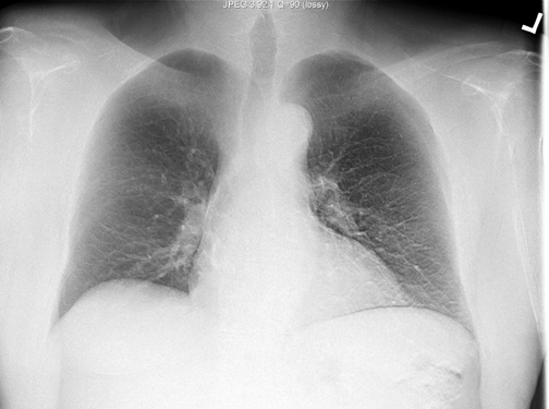
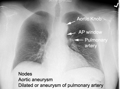
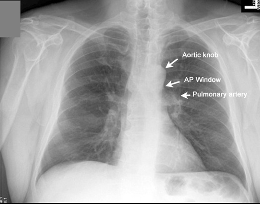
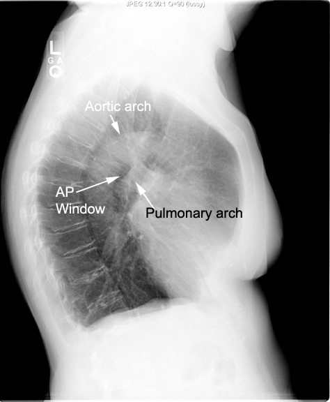
| CXR | Identify Aortic knob, Aortopulmonary window (AP window) and the main Pulmonary artery | What is the significance of full AP window? | Answer |
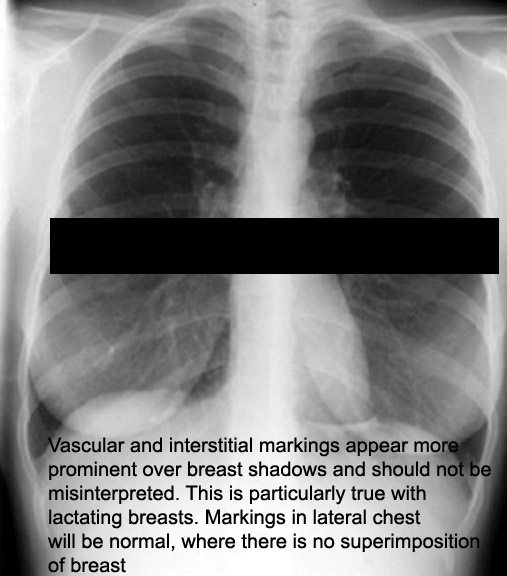
| CXR | Identify breast shadow | What is its significance of breast shadow in evaluating lower lung fields? | Answer |
| What is the significance of one missing breast? | Answer | ||
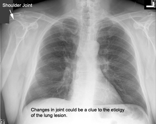
| CXR | Identify shoulder joint | Why is it important to look at shoulder? | Answer |
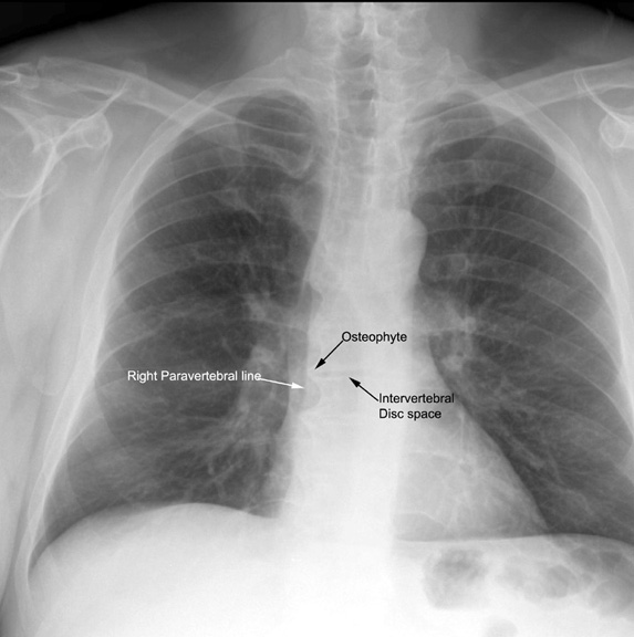
| CXR | Identify right para vertebral line . | What are the conditions that can give prominent right para vertebral line? | Answer |
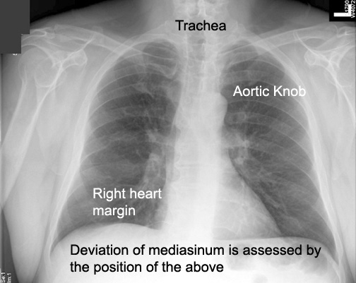
| CXR | Hpw do you judge position of mediastinum? | Answer | |
|
Lobar Projections |
|||
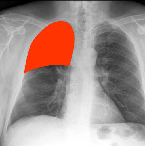
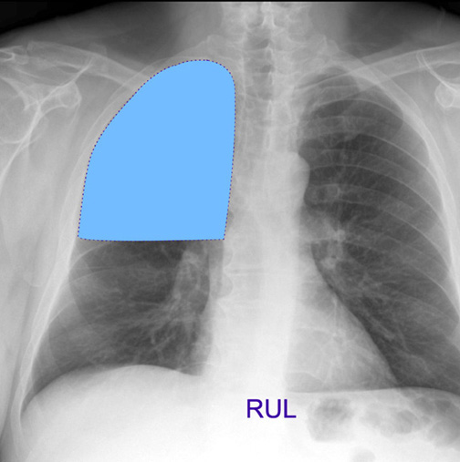
| CXR | RUL projection in PA view | Answer | |
| CXR | RUL projection in PA view | Answer | |
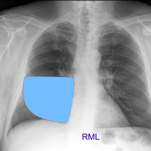
| CXR | RML projection in PA view | Answer | |
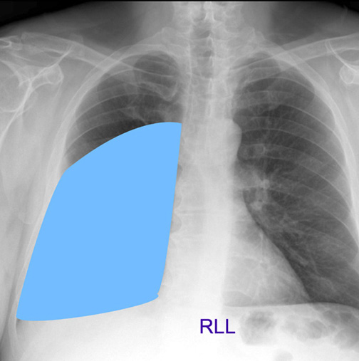
| CXR | RLL projection in PA view | Answer | |
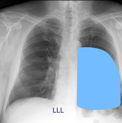
| CXR | LLL projection in PA view | Answer | |
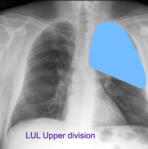
| CXR | LUL projection in PA view | Answer | |
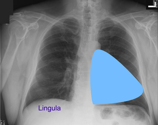
| CXR | Lingula projection in PA view | Answer | |
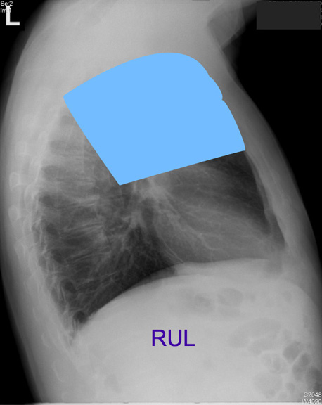
| CXR | RUL projection in lateral view | Answer | |
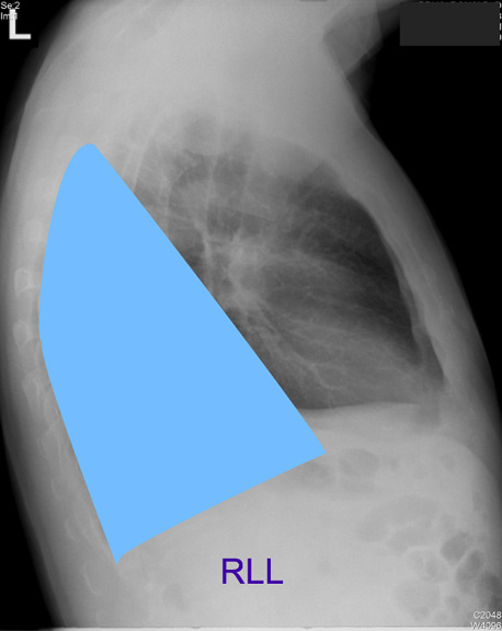
| ICXR | RLL projection in lateral view | Answer | |
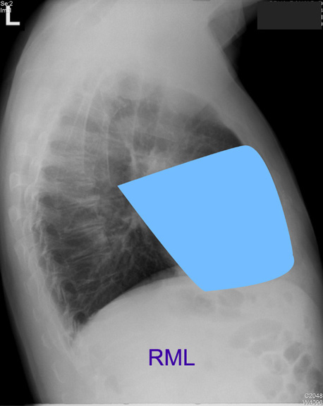
| CXR | RML projection in lateral view | Answer | |
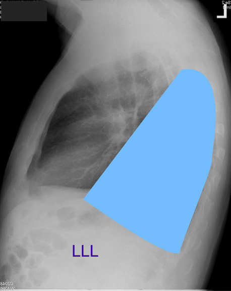
| CXR | LLL projection in lateral view | Answer | |
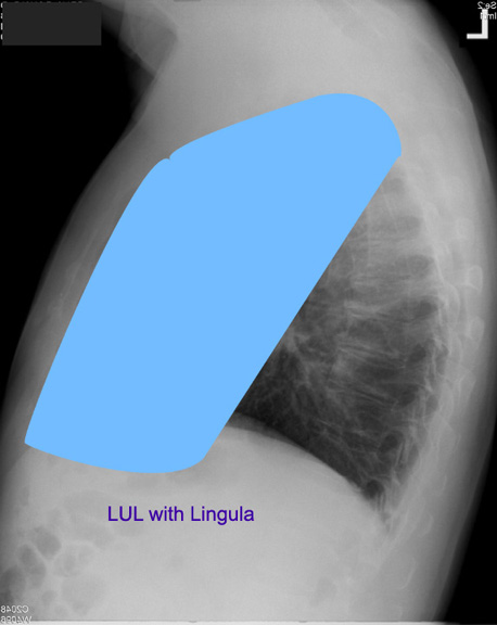
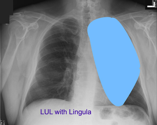
| CXR | LUL with lingula projection in lateral view | Answer | |
| CXR | LUL with lingula projection in PA view | Answer | |
|
Tracheo Bronchial Tree |
|||
| Drawing | Identify the major divisions of the Tracheobronchial tree. | ||
| Bronchogram | |||
| Bronchogram | Locate the Carina | ||
| Bronchogram | Identify the divisions of left main bronchus | ||
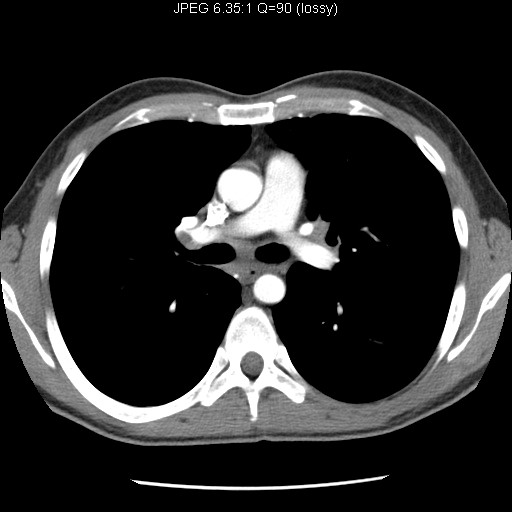
| CT | Identify the bronchus | ||
| CT | Identify the labeled bronchi | ||
| CT | Identify the bronchus | ||
| CT | Identify the labeled bronchi | ||
| CT | Identify the labeled bronchi | ||
| CT | Identify the labeled bronchi | ||
| CT | Identify the labeled bronchi | ||
| CT | Identify the labeled bronchi | ||
| CT | Identify the labeled bronchi | ||
|
Heart |
|||
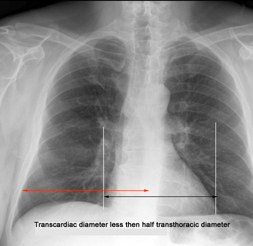
| CXR | How do you measure heart size? | What is the normal range for heart size? | Answer |
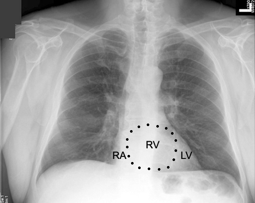
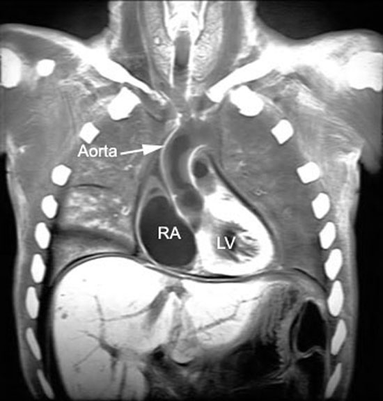
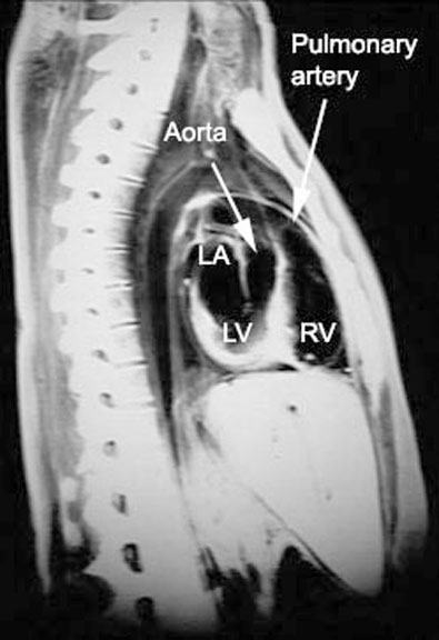
| CXR | Identify chambers of heart in PA view | Answer | |
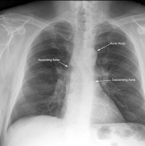
| CXR | Identify ascending aorta, aortic knob and descending aorta | Answer | |
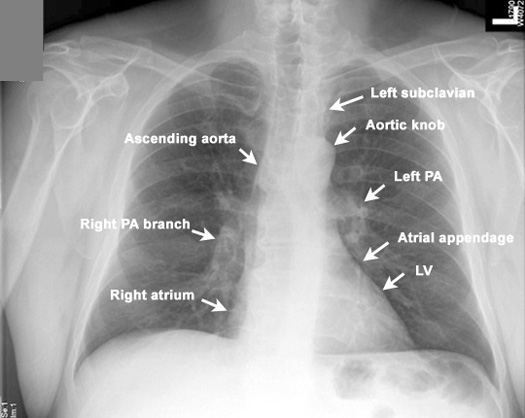
| CXR | Identify structures along the edge of mediastinum | Answer | |
| CXR | Identify Aortic knob, AP window and Pulmonary artery | What is the significance of full AP window? | Answer |
|
Lateral view |
|||
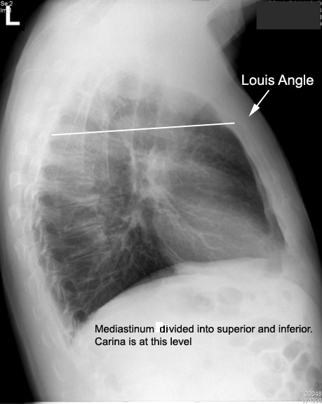
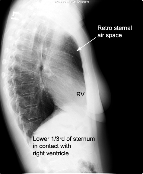
| CXR | Identify Sternum and angle. of Louis. | What are the important landmarks at Sternal angle (Angle of Louis)? | Answer |
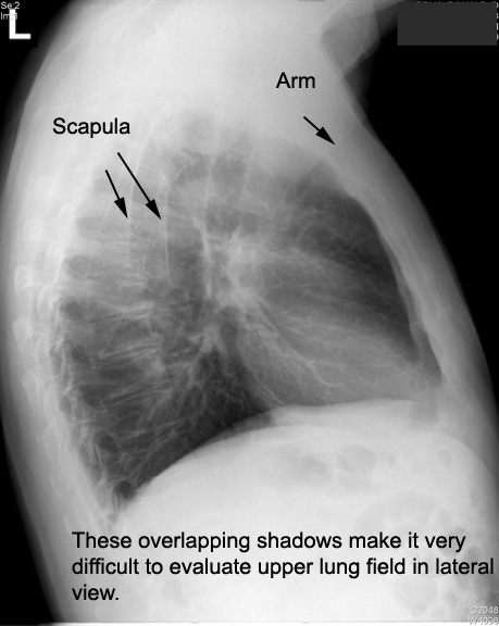
| CXR | Identify axillary fold and Scapula | Answer | |
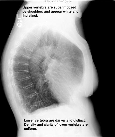
| CXR | IIdentify Vertebra. | Is there a difference in radio density along the Spine. | Answer |
| CXR | Identify aortic, pulmonary arches and AP window | Answer | |
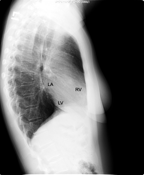
| CXR | Identify chambers of heart in lateral view | Answer | |
| CXR | Identify retro sternal air space. | What is normal retro sternal space? | Answer |
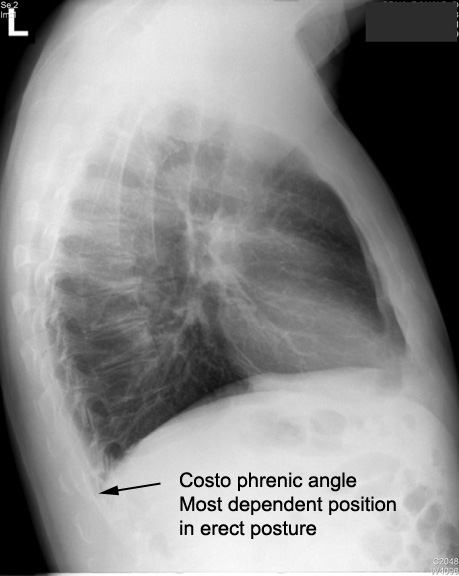
| CXR | Identify costophrenic angle | How do we use this information? | Answer |
| CXR | Identify oblique and transverse fissure | What is the normal position of these fissures? | Answer |
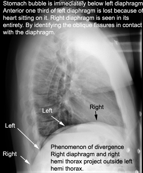
| CXR | Identify diaphragms left and right | How did you arrive at your answer? | Answer |
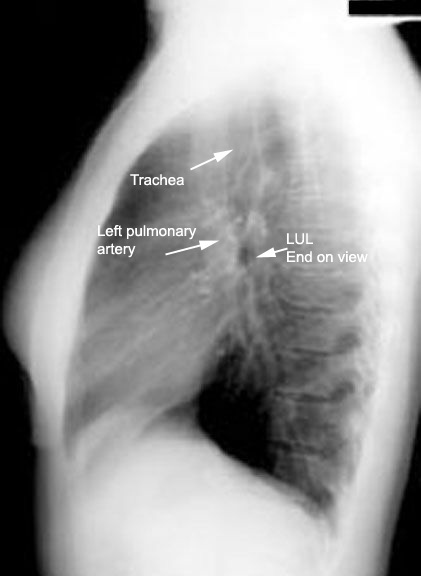
| CXR | Identify trachea and LUL orifice. | Answer | |
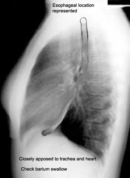
| CXR | Identify the position of esophagus. | Answer | |
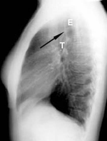
| CXR | Identify Trcheo esophageal stripe | How do we use this information? | Answer |
| CXR | Identify left and right pulmonary artery | Answer | |
|
CT |
|||
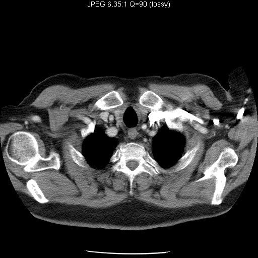
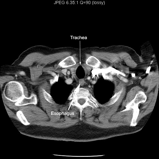
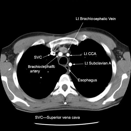
| Image 1 | Identify Trachea and Esophagus | Answer | |
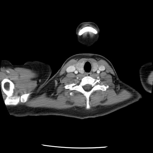
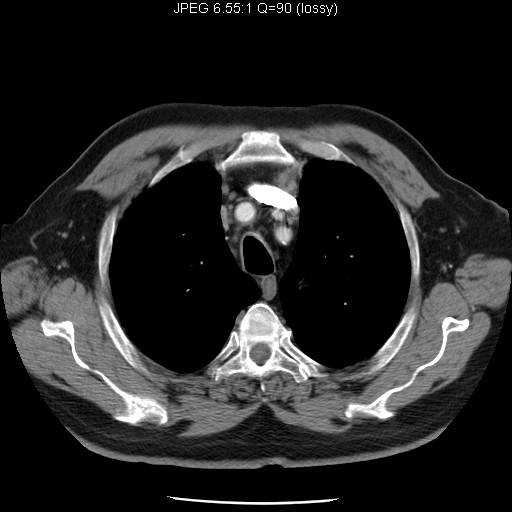
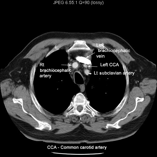
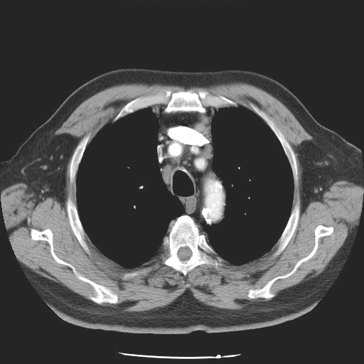
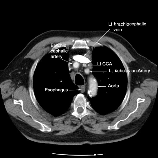
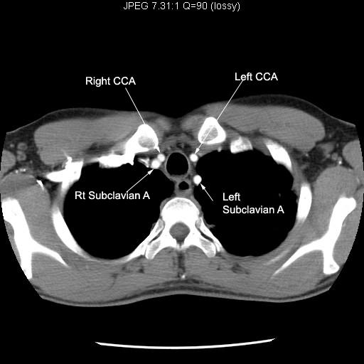
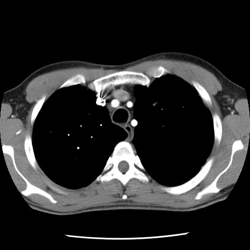
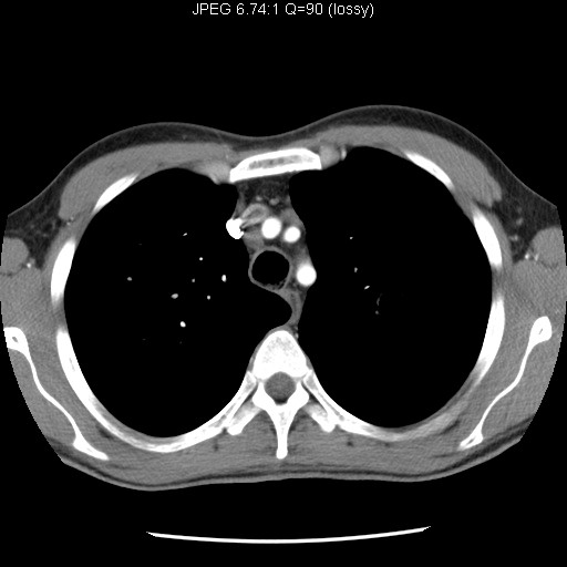
| Image 2 | Identify vascular structures | Answer | |
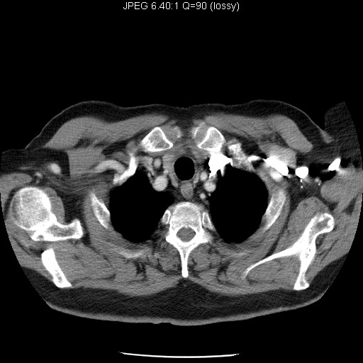
| Image 3 | Identify vascular structures | Answer | |
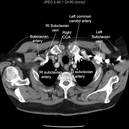
| Image 4 | Identify vascular structures | Answer | |
| Image 5 | Identify vascular structures | Answer | |
| Image 6 | Identify vascular structures | Answer | |
| Image 7 | Identify vascular structures | Answer | |
| Image 8 | Identify vascular structures | Answer | |
| Image 9 | Identify vascular structures | Answer | |
| Image 10 | Identify vascular structures | Answer | |
| Image 11 | Identify vascular structures | Answer | |
| Image 12 | Identify vascular structures | Answer | |
| Image 13 | Identify vascular structures | Answer | |
| Image 14 | Identify vascular structures | Answer | |
| Image 15 | Identify vascular structures | Answer | |
| Image 16 | Identify vascular structures | Answer | |
|
CT |
|||
| Major veins
|
Sequence1
Sequence2 Sequence3
Sequence4 Sequence5
Sequence6 Sequence7
Follow the major veins draining into superior vena cava. |
||
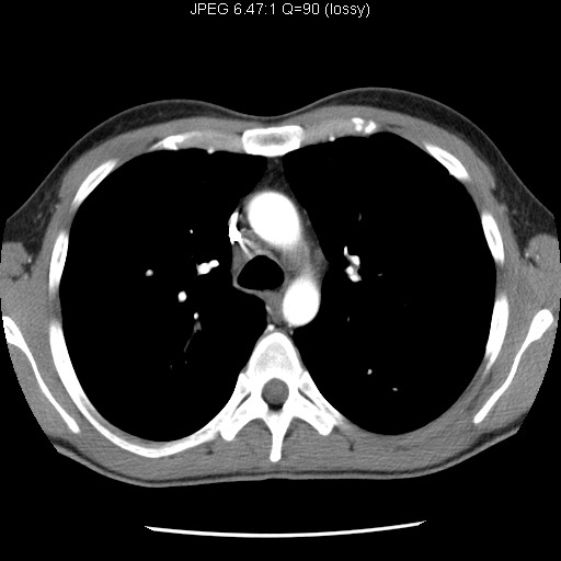
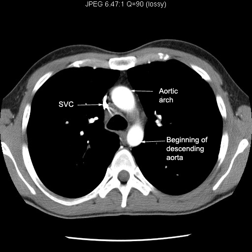
| SVC | Right and left brachiocephalic veins join to form superior vena cava. Follow the superior vena cava entering right atrium | ||
| Right atrium | Sequence
1 Sequence 2 Sequence
3 Sequence 4
Follow SVC entering right atrium. Note the relationship of right atrium to other chambers of heart. |
||
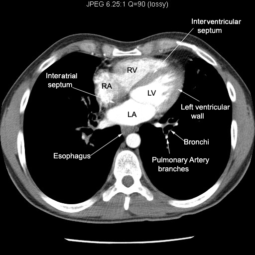
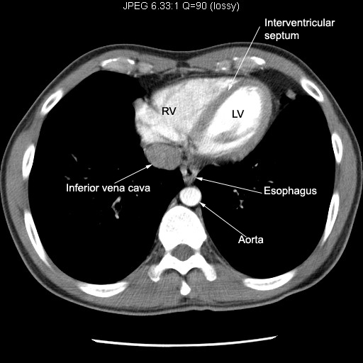
| Right ventricle | Sequence 1 Sequence 2 Sequence 3 Sequence 4 Note the relationship of right ventricle to Sternum. |
||
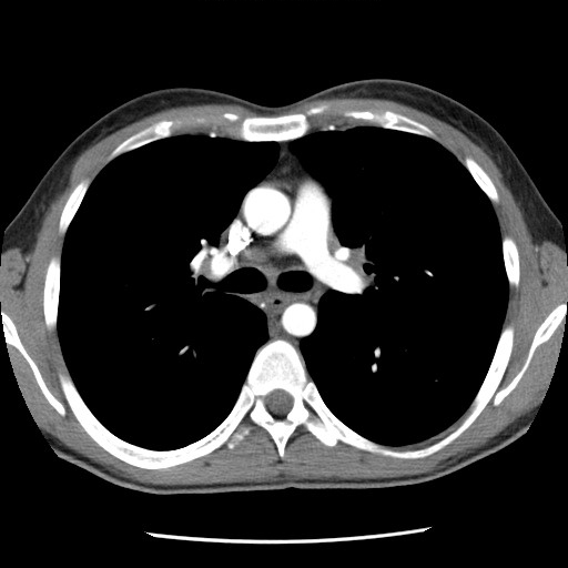
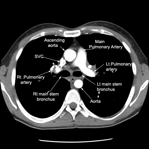
| Pulmonary artery |
Follow pulmonary artery. Identify right and left branches. |
||
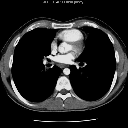
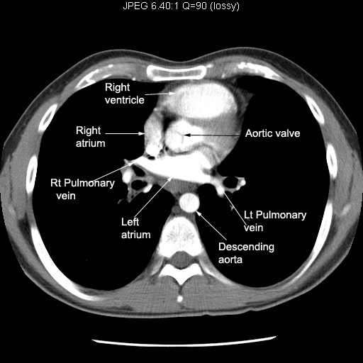
| Pulmonary veins | Sequence 1 Sequence 2 Sequence 3 Note Pulmonary veins entering left atrium. |
||
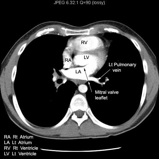
| Left atrium | Sequence 1 Sequence 2 Sequence 3 Note mitral valve leaflet. Note its location.. |
||
| Left ventricle |
Note left ventricular wall thickness and Inter ventricular septum |
||
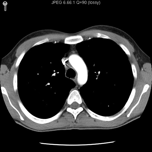
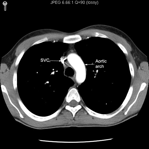
| Aorta | Sequence 1 Sequence 2 Sequence 3 Sequence 4 Sequence 5 Sequence 6 Sequence 7 Sequence 8 Follow Aorta from head end to foot. Note major branches, ascending, arch and descending portions of Aorta. |
||
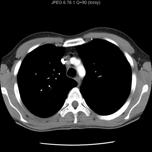
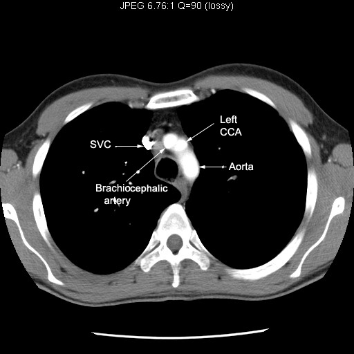
| Major branches | Sequence
1 Sequence 2 Sequence
3 Sequence 4
Follow the major branches of Aortic arch to neck. |
||