The images are not necessarily from this case.
RSV Pneumonia
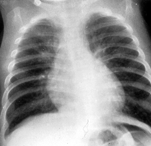
Chest x-ray 1 There
are mild peribronchial infiltrates and the lungs are hyperinflated because of
air trapping (air gets in but can’t get out ) because of mucous necrotic
debris in the airways.
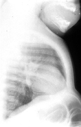
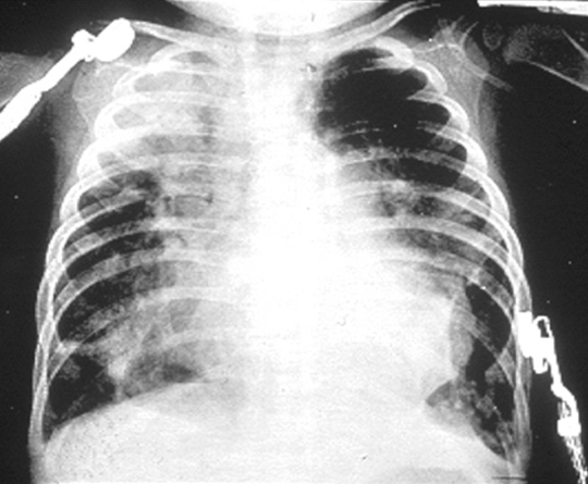
Chest x-ray 2
Shows
that the lungs are hyperinflated, because diaphragms are flattened.
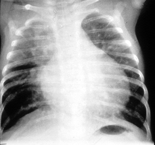
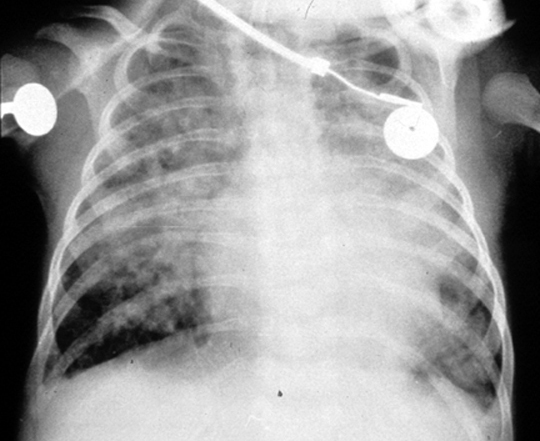
CXR 3-5: Increasingly progressive RSV pneumonia in infants with congenital heart disease. There are patchy infiltrates and hyperinflation from air trapping. The presence of infiltrates is common in severe RSV pneumonia and does not imply the presence of a secondary bacterial pneumonia.
Send comments to Dr. A.J. Chandrasekhar M.D.