The images are not from this case.
Blood smear in a patient with bacterial infection:
Doehle bodies: Discrete round or oval density in the periphery of cytoplasm. Stains sky blue with Romanowsky stain
Staphylococcus Aureus
Staph aureus/Pneumonia. (Dr Aliya Husain)
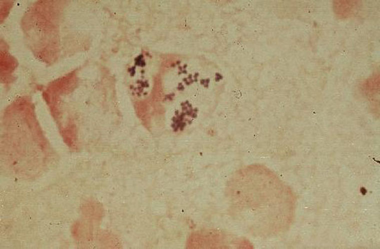
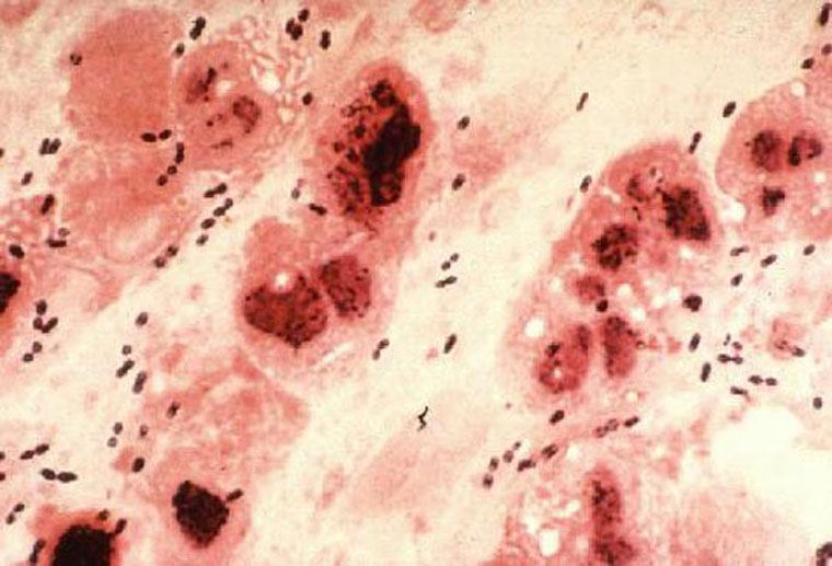
Staphylococcus Aureus: a gram stain of a smple of fluid from a skin lesion reveals clusters of gram positive cocci. Several neutrophiles are noted.
(Dr Ralph Leischner)
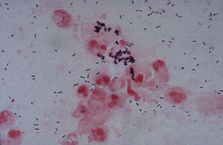
Sputum smear Staphylococcus & Streptococcus pneumoniae
(Dr Tadayo Hashimoto)
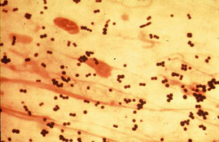
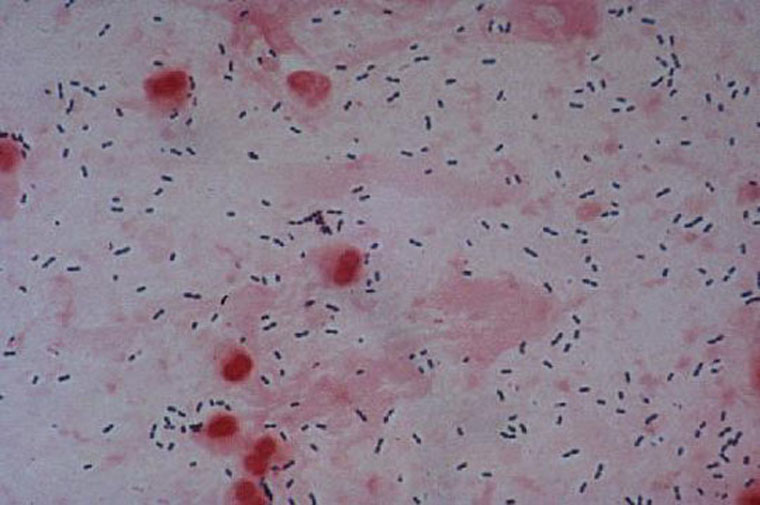
Sputum smear Staphylococcus aureus (Dr Tadayo Hashimoto)
Pus smear (wound) Staphylococcus aureus (Dr Tadayo Hashimoto)
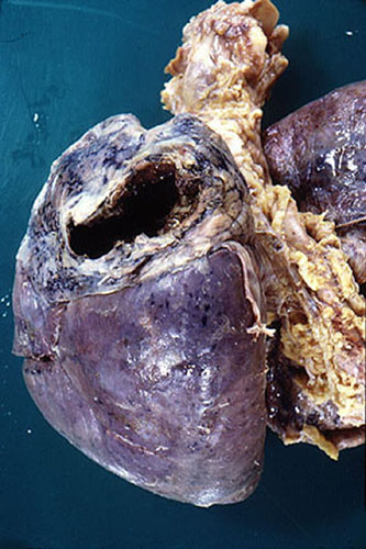
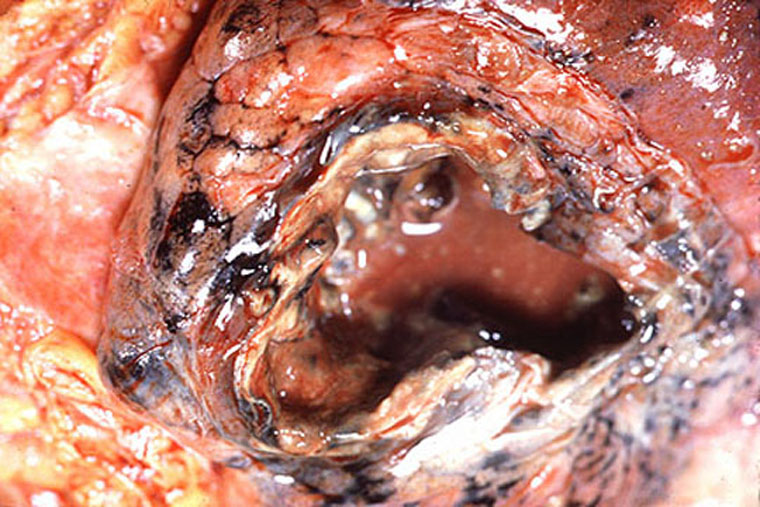
Lung abscess /gross. (Dr Ralph Leischner)
Lung abscess/ gross. (Dr Ralph Leischner)
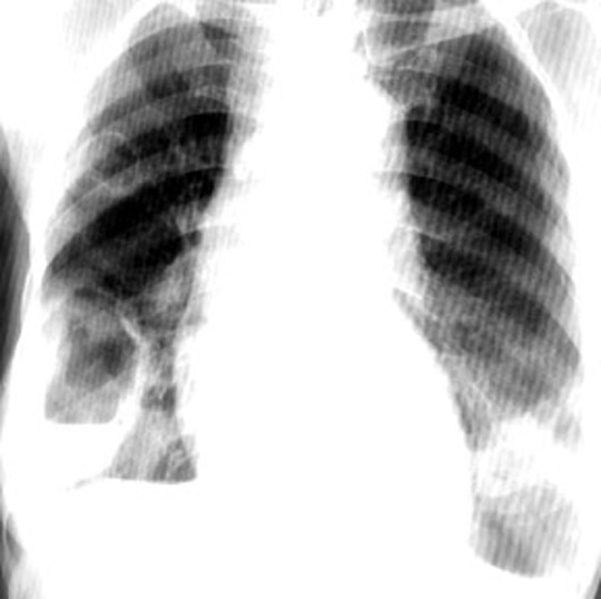
CXR - Lung abscess (Dr Arcot Chandrasekhar )
Streptococcus Pyogenes:
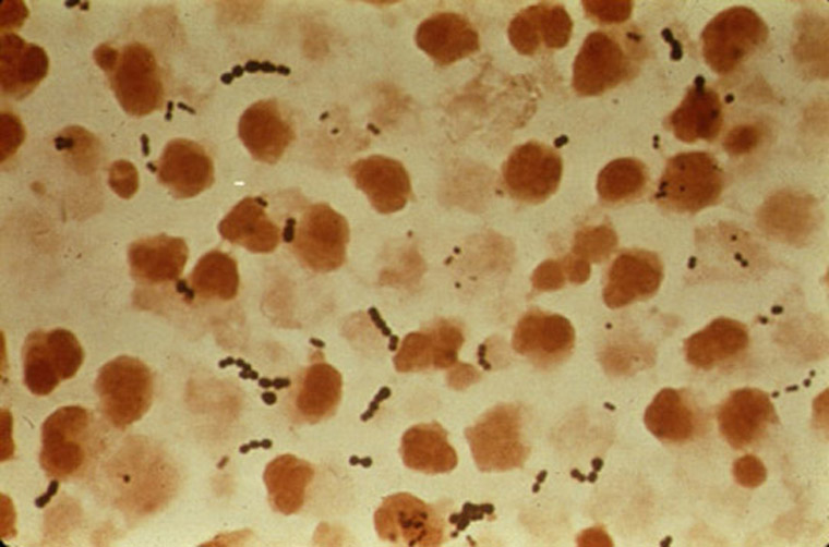
Streptococcus pneumonia: a grm stain of sputum reveals gram positive diplococci intermixed with neutrophils and mucin. (Dr Ralph Leischner)
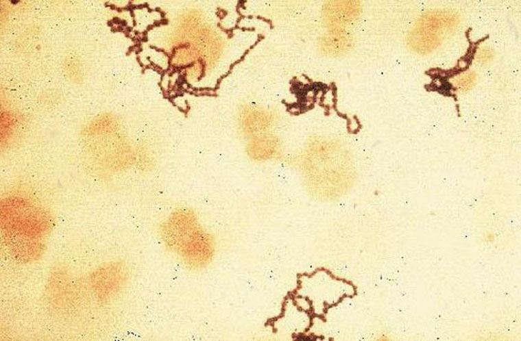
Streptococcus Pyogenes: A gram stain of a sample of body fluid reveals long and short chains of gram positive cocci. The bacteria are intermixed with neutrophiles. (Dr Ralph Leischner)
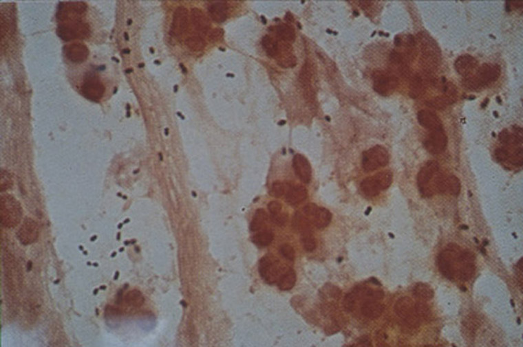
Sputum smear Streptococcus pneumoniae pneumoniae (Dr Tadayo Hashimoto)
Sputum smear Streptococcus pneumoniae/PMN pneumoniae (Dr Tadayo Hashimoto)
Knee tap smear Group A, Streptococcus pyogenes (Dr Tadayo Hashimoto)
Sputum smear Streptococcus pneumoniae pneumoniae (Dr Tadayo Hashimoto)
Streptococcus Pyogenes: A gram stain of a sample of body fluid reveals long and short chains of gram positive cocci. The bacteria are intermixed with neutrophiles. (Dr Ralph Leischner)
Streptococcus pneumonia: a gram stain of sputum reveals gram positive diplococci intermixed with neutrophils and mucin. (Dr Ralph Leischner)
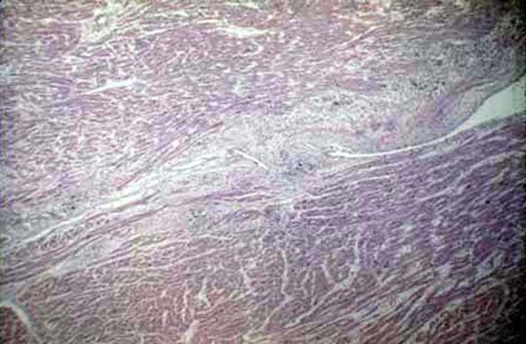
Rheumatic fever
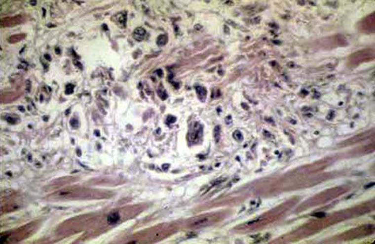
Acute Rheumatic heart disease (Dr Ralph Leischner)
Acute Rheumatic heart disease (Dr Ralph Leischner)
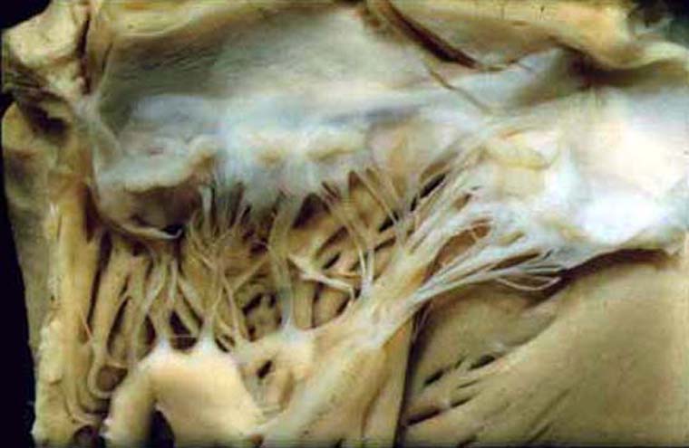
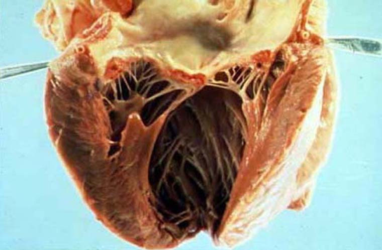
Acute Rh valvular heart disease/vegetations on inflamed mitral valve leaflets. (Dr Ralph Leischner)
Mitral regurgitation. Murmur (Dr Robert Lichtenberg)
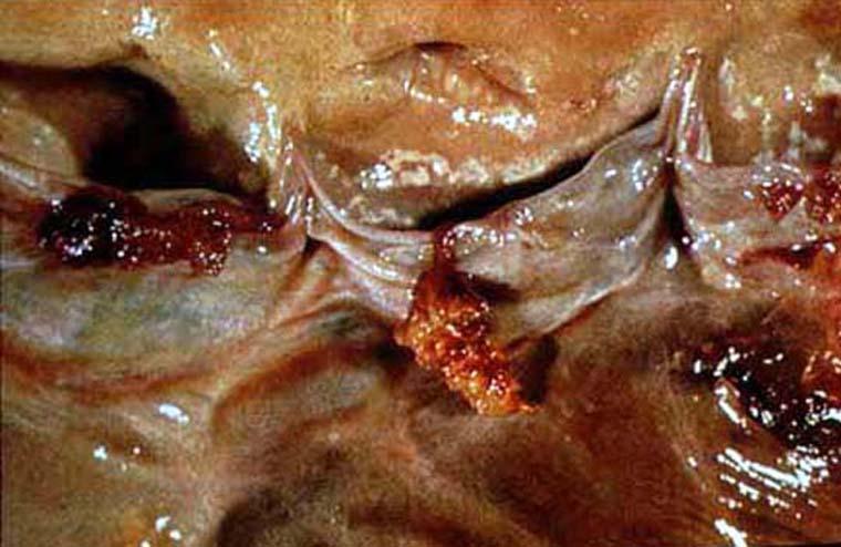
Endocarditis
Infective endocarditis/Vegetations adherent to valve (Dr Ralph Leischner)
Endocarditis/vegetations (Dr Ralph Leischner)
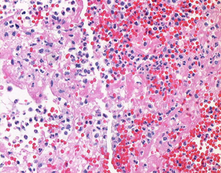
Infective Endocarditis Renal Infarct/Histology/40 (Dr Ralph Leischner). This is a an embolic complication.
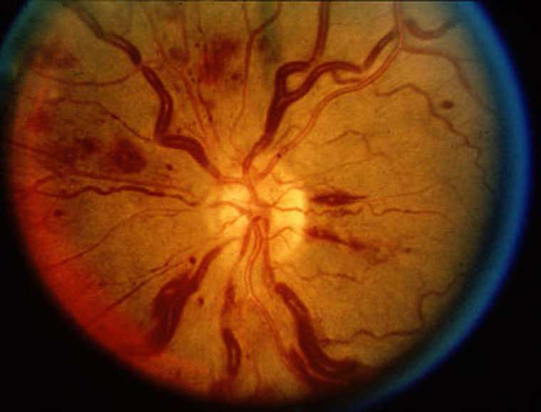
Tortousity of retinal veins and hemorrhage due to macroglobulinemia (Dr Harry Messmore) This image is just to show you retinal hemorrhage. In Roth spots the center of hemorrhage will be white.
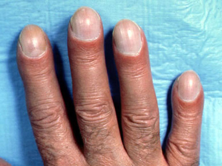
Clubbing of fingers in patient with chronic pulmonary disease and lung disease (Dr Arcot Chandrasekhar ). This is just an example to show you what the mild clubbing of SBE will look like.
Echocardiogram
Send comments to Dr. A.J. Chandrasekhar M.D.