The images are not necessarily from this case.
Case 1
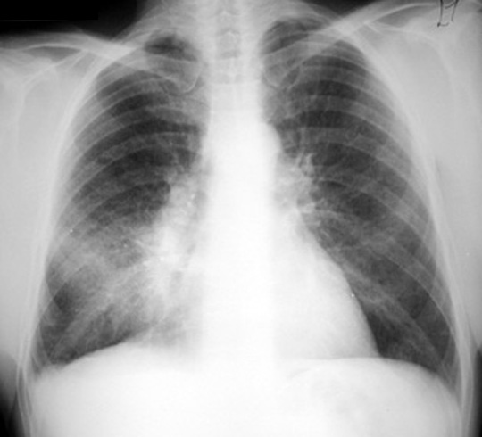
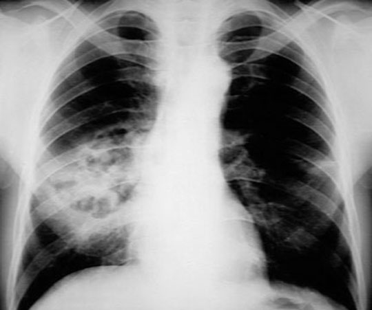
CXR Case 1: Density in the projection of right mid lung field silhouetting with right heart margin suggesting RML disease. Note also right hilar fullness.
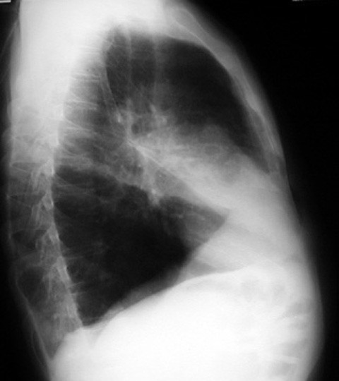
CXR Lateral: Consolidation in the projection of RML.
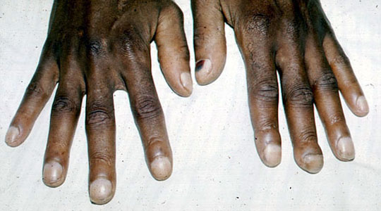
Clubbing: Bulbous swelling of distal fingers from a patient with lung cancer.
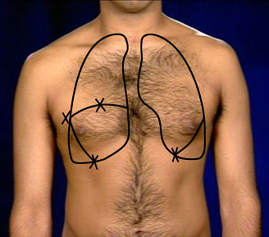
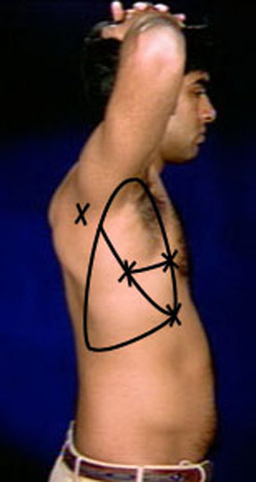
Surface anatomy lobes: To demonstrate the location of findings corresponding to RML
Surface anatomy lobes: To demonstrate the location of findings corresponding to RML
Bronchial breathing with crackles: Note the pitch, expiration as long as inspiration with a pause in between.
Community acquired pneumonia: List of organisms
Host predisposing factors: List of defects
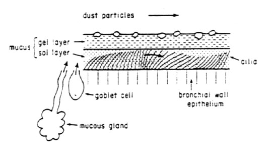
Pulmonary defense drawing
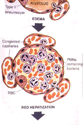
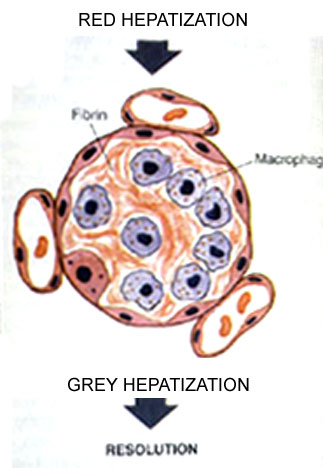
Lobar pneumonia: Note the lobar distribution.
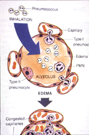
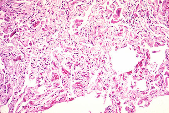
Histology: alveoli filled with inflammatory cells
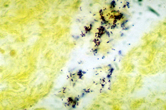
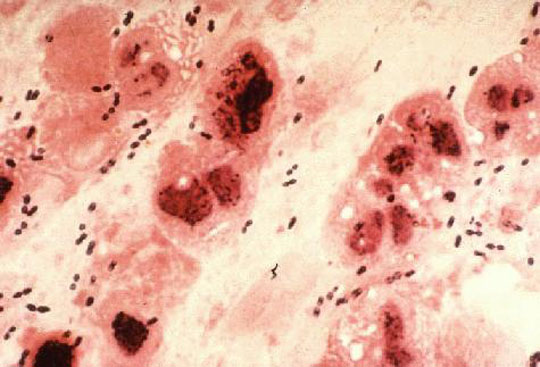
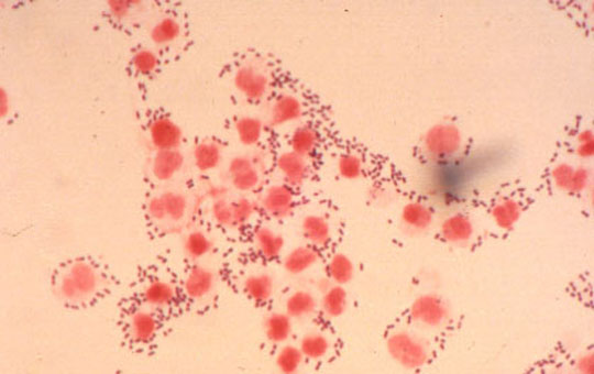
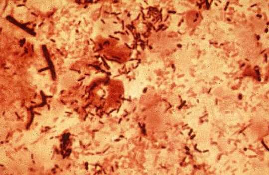
Pneumococcus: Gram positive diplococci
Pneumococus: Gram positive diplococci
Pneumococcus: Gram positive diplococci
Pneumococcus: Gram positive diplococci
Case 2
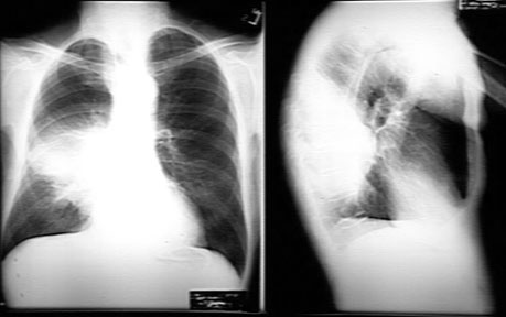
Case 2 CXR: Lateral showing patchy infiltrates
Bullous myringitis
Atypical pneumonia/ agents: List of organisms
Mycoplasma pneumoniae
Histology
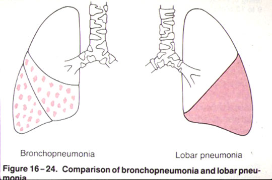
Bronchopneumonia: Note patchy infiltrates
Lobar/broncho pneumonia: Diagrammatic representation
Viral pneumonia micro: Lymphoctic infiltration of interstitium.
Case 3
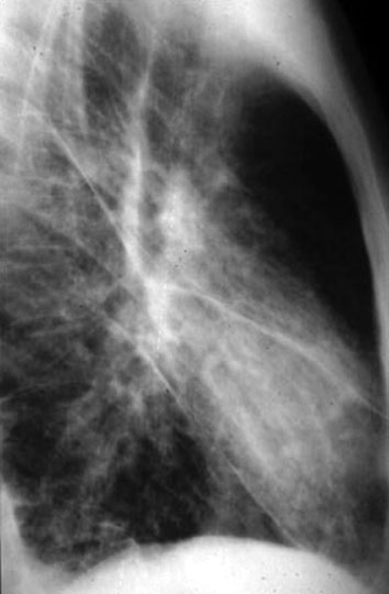
CXR initial: Consolidation of superior segment of RLL
CXR Proceeding to necrosis and cavitations
CXR follow up PA
CXR follow up lateral
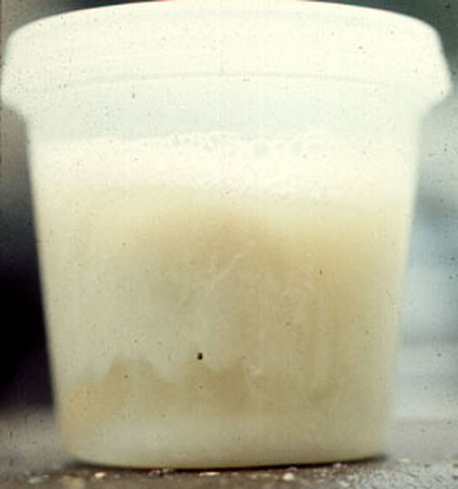
Sputum: 3 layered sputum (Purulent sediment, clear liquid, foam on top)
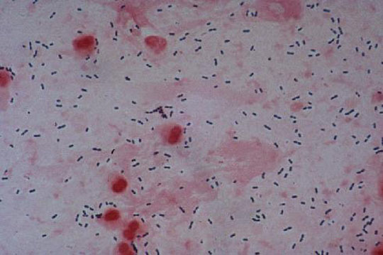
Gram stain? peritoneal fluid: Mixed organisms
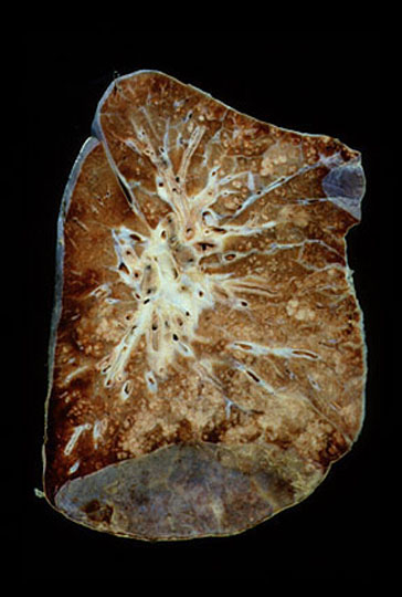
Lung abscess path: Necrotic lung tissue
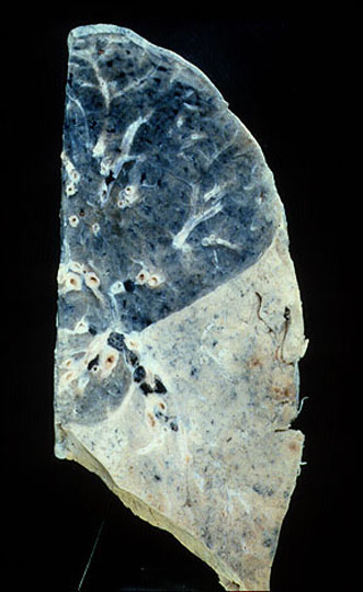
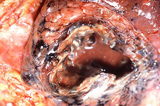
Gross lungs: Hole in lung
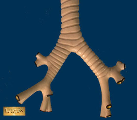
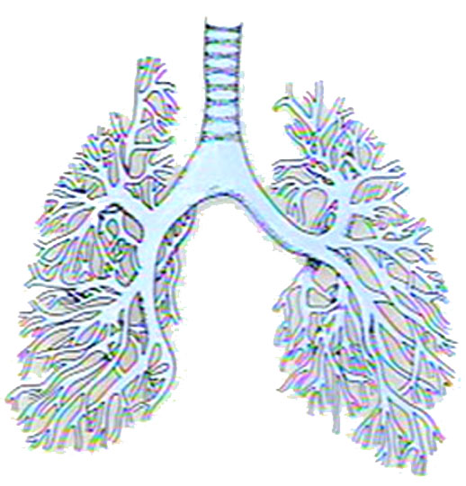
Bronchial tree Model
Drawing Bronchial tree
Clubbing: Bulbous swelling of fingers in a patient with metastatic lung disease
Send comments to Dr. A.J. Chandrasekhar M.D.