Learning Objectives: You should be able to:
- Draw a generalized diagram of how information flows to and
from the central nervous system for the state-dependent regulation of
breathing.
- List the paticipant nuclei in the brain stem responsible
for the rhythmic and coordinated act of automatic breathing.
- Identify ten mechanoreceptor reflexes that alter the
respiratory pattern to overcome various perturbances to normal breathing.
- Explain the protective role of peripheral and central
chemoreceptors in normal breathing and pathological situations.
Rhoades & Tanner Text Readings: Chapter 22, Pages 401-414
Neural Organization
Respiratory Controller
Mechanoreflexes
Chemoreflexes
MainMenu
General Neural Organization
- Overview
- breathing is a coordinated act which is subjected to
many feedforward and feedback controls
- the system has a hierarchical neural organization with
involuntary and voluntary components
- PaCO2 seems to be the regulated variable, but blood gas
values of carbon dioxide fluctuate widely within normal, healthy
individuals
- Major Components of the Respiratory System
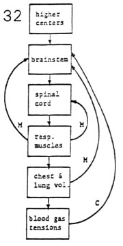
- respiratory controllers
- higher center feed-forwards (voluntary)
- brainstem (automatic)
- spinal cord 32
- respiratory effectors
- respiratory muscles
- chest wall and lung
- respiratory outputs
- alveolar ventilation
- blood gas tensions
- respiratory sensors
- mechanical feedbacks
- chemical feedbacks
- Cortical Influences
- phonation and articulation
- maximum voluntary ventilation
- breathholding
- central hypoventilation syndrome ("Ondine's
Curse")
- failure of automatic respiratory control when
asleep
- auxiliary ventilatory support is usually indicated
- Abnormal Respiratory Patterns (Fig. 33)
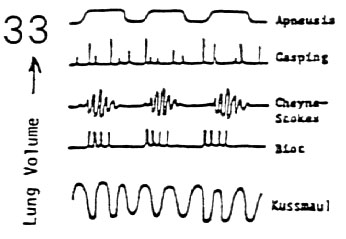
- mechanical or vascular damage to the CNS can result in
ominous breathing patterns
- apneusis: rostral pontine damage
- gasping: extensive pontine damage, severe hypoxia
- Cheyne-Stokes respiration (spindle pattern): poor
brain stem perfusion
- Biot breathing (cluster pattern): pontine
malfunction
- Kussmaul: metabolic acidosis
- apnea: high cervical cord or extensive medullary
damage
Neural Organization
Respiratory Controller
Mechanoreflexes
Chemoreflexes
MainMenu
Central Respiratory Controller
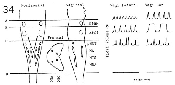
- Nucleus Tractus Solitarius (NTS, dorsomedial medulla)
- I responsible for phrenic motoneuron activation
- Iß responsible for vagal afferent integration
- Nucleus Ambiguus (NA, ventrolateral, middle medulla)
- motor I cells that activate upper airway musculature
(larynx, pharynx)
- Nucleus Retroambigualis (NRA, ventrolateral, caudal
medulla)
- premotor I cells (rostral) that activate external
intercostal motoneurons
- premotor E cells (caudal) that activate internal
intercostal motoneurons
- Nucleus Parabrachialis Medialis (NPBM, dorsolateral pons)
- tonic discharging cells inhibitory to Ià cells of
inspiration
- commonly known as the pneumotaxic center
- Pre-Bö tzinger Complex (pBOT, ventrolateral, rostral
medulla)
- pacemaker I neurons responsible for generating
respiratory rhythm
Neural Organization
Respiratory Controller
Mechanoreflexes
Chemoreflexes
MainMenu
Mechanoreflexes and Breathing
- Vagal reflexes from the lungs
- slowly adapting pulmonary stretch receptors
- location: nerve filaments in smooth muscles of
trachea and bronchi
- stimulation: lung inflation (static volume
detector)
- reflex response: termination of inspiration (Hering-Breuer
reflex)
- rapidly adapting irritant receptors
- location: unmyelinated nerve filaments in the
epithelium of larynx, trachea, & extra pulmonary bronchi
- stimulation: noxious irritants, distortion of
larynx, large lung inflations & deflations, pneumothorax
- reflex responses: cough, hyperventilation,
bronchoconstriction
- pulmonary C-fiber receptors
- location: unmyelinated fibers in the alveolar wall
- stimulation: vascular engorgement, interstitial
edema fluid formation
- reflex response: rapid shallow breathing, large
airway constriction, bradycardia
- bronchial C-fiber receptors
- location: unmyelinated fibers in blood vessel walls
of the bronchial circulation
- stimulation: vascular congestion, chemical injury,
microemboli
- reflex responses: like pulmonary C-fiber
receptor responses, sensation of dyspnea?
- Reflexes from the Upper Airways
- nasal passage receptors
- location: nasal air passages
- afferent pathway: trigeminal nerve
- stimulation: mechanical or chemical irritation
- reflex response: sneeze, bradycardia
- larynx and tracheo-bronchial receptors
- location: large upper airways
- afferent pathway: vagus nerve, superior laryngeal
nerve
- stimulation: mechanical or chemical irritation
- reflex response: cough, wide swings in blood
pressure, Valsalva
- pharynx receptors
- location: receptors in the walls of the common
air-food passageway
- afferent pathway: vagus nerves, glossopharyngeal
nerves
- stimulation: peristaltic wave during swallowing
- reflex response: closure of glottis
- other reflexes of unknown origin
- hiccup (dry food, misplaced cardiac pacemaker)
- yawning (sleepiness, boredom, intracranial disease)
- sighing (periodic deep inspiration every 15 to 30
sec)
- Spinal Cord Reflexes
- muscle spindle receptors (somatic afferents to thoracic
spinal cord
- location: intercostal musculature (very few muscle
spindles in diaphragm)
- afferent pathway: somatic afferents to thoracic
spinal cord
- stimulation: low total compliance
- reflex response: additional respiratory muscle
activation to overcome low compliance
- nociceptive pain receptors
- location: bare nerve endings in viscera, muscles,
bone
- afferent pathway: visceral and somatic afferents
- stimulation: ischemia, bodily injury
- reflex response: gasp, excited breathing,
hypoventilation, apnea
- joint receptors
- location: joints of arms, legs, feet
- afferent pathway: somatic afferents to spinal cord
- stimulation: joint motion in exercise
- reflex response: increased frequency and volume of
breathing
Neural Organization
Respiratory Controller
Mechanoreflexes
Chemoreflexes
MainMenu
Chemoreflexes and Breathing
- Location of Chemoreceptors
- chemoreceptors are located exclusively on the arterial
side of the circulation
- peripheral chemoreceptors: aortic and carotid
bodies sense
- central chemoreceptors: chemosensitive zone of
medulla sense
- there are no venous chemoreceptors that
"taste" the venous blood
- there are no airway chemoreceptors that
"smell" the alveolar air
- CO2 Response Curve (Fig. 35)
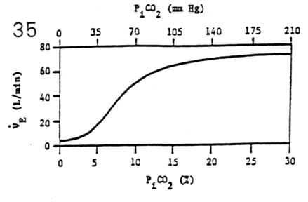
- minute volume (V E = frequency (f) * tidal volume (VT)
- V E is a sigmoidal function of inspired PiCO2
- CO2 sensitivity = slope of plot over 5%-7% range (6
L/min per %CO2)
- CO2 sensitivity decreases in sleep and presence of
drugs
- maximal CO2 responsivity is about 60% of maximum
voluntary ventilation
- O2 Response Curve (Fig. 36)
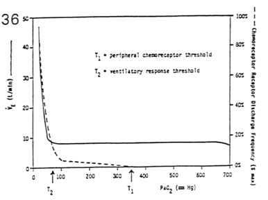
- minute volume (V E) = frequency (f) * tidal volume (VT)
- V E is a hyperbolic function of inspired PiO2 (mm Hg)
- O2 response threshold: region where V E increases more
steeply with
- � PaO2 (÷70 mm Hg)
- normal PaO2 of 95 mm Hg is below its threshold
(indirectly regulated)
- O2 chemoreceptor threshold: region where receptors
start discharging
- with � PaO2 (÷350 mm Hg)
- receptors are discharging at PaO2 of 95 mm Hg, but
they have no ventilatory effect
- Ventilation and Integration of Chemoreceptor Inputs
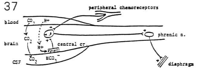
- respiratory neurons of the brainstem possess no
inherent chemosensitivity to low O2
- ventilatory outputs depend upon intact central and
peripheral chemoreceptor afferent projections to respiratory nuclei of
medulla and pons
- peripheral chemoreceptors primarily respond to low PaO2
(< 70 mm Hg)
- aortic bodies and carotid bodies
- peripheral chemoreceptors can respond secondarily
to � PaCO2 and � [H+]
- central chemoreceptors can respond to elevated PaCO2
and/or elevated [H+]
- central receptors are embedded within ventrolateral
surface of medulla
- CO2 crosses blood-brain barrier very quickly
- H+ crosses blood-brain barrier very slowly
- cerebral spinal fluid buffers response (pH = 7.32)
- central and peripheral chemoreceptor inputs mutually
potentiate each other
- peripheral chemoreceptors protect the central nervous
system against global hypoxia and neuronal depression
Neural Organization
Respiratory Controller
Mechanoreflexes
Chemoreflexes
MainMenu


