|
This has two modules
UNDER CONSTRUCTION
|
|
| This exercise is for students studying Anatomy. Try answering the questions from the unlabeled images first. Click on the images to see the labeled image. | |
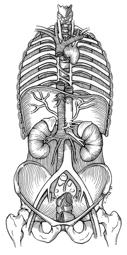
| Diagram | IVC and Aorta |
| Course | |
| Reconstructed Image | Where does Abdominal Aorta start and end . |
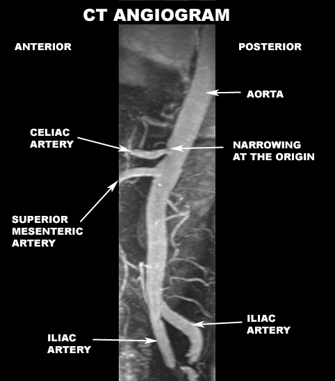
| CT | Distal Aorta |
| CT | Aorta is round |
| CT | Aorta is oval just before bifurcation |
| CT | Aorta has bifurcated |
| CT | Aortic Bifurcation |
| Branches | |
| PA view Reconstructed Image | Identify the branches of Abdominal Aorta |
| Lateral view Reconstructed Image | Identify the branches of Abdominal Aorta |
| CT | Identify Lumbar arteries |
| Relationship | |
| CT | Proximal aorta relationships |
| CT | Proximal aorta relationships |
| CT | Pancreas is anterior to Aorta |
| CT | Note that the left renal vein crossing anterior to Aorta |
| Appearance in | |
| Color Doppler | Aorta |
| MRI | Aortic Bifurcation |
| US | Aorta |
| US | Aortic bifurcation |
| Course | |
| PA view Reconstructed image | Identify Celiac artery |
| Lateral view Reconstructed image | Identify Celiac artery |
| Lateral view Reconstructed image | Celiac artery |
| CT | Origin of Celiac artery |
| Branches | |
| Reconstructed image | Identify branches of Celiac artery |
| CT | Identify branches of Celiac artery |
| CT | Identify branches of Celiac artery |
| Case | Gastro duodenal artery a branch of Hepatic artery |
| Relationship | |
| Appearance in | |
| US | Celiac artery with splenic and hepatic artery branches |
| US | Hepatic Artery |
| US | Hepatic Artery |
| Color Doppler | Celiac artery with splenic and hepatic artery branches |
| Drawing or CT | What organs are supplied by Celiac artery |
| Course | |
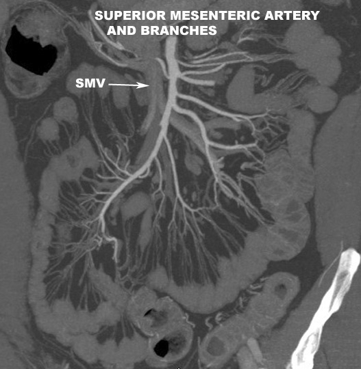
| PA view Reconstructed image | Identify Superior Mesenteric artery |
| Lateral view Reconstructed image | Identify Superior Mesenteric artery |
| Lateral view Reconstructed image | Identify Superior Mesenteric artery |
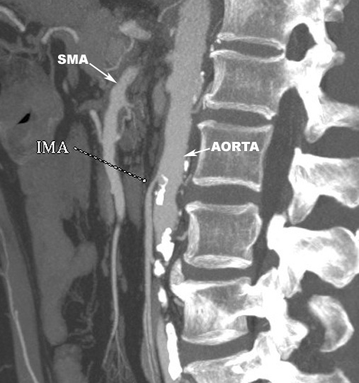
| CT | Origin of SMA |
| Branches | |
| PA view Reconstructed image | Branches of SMA |
| Relationship | |
| CT | Behind Pancreas |
| CT | Anterior to Uncinate process |
| CT | Relationship to structures |
| CT | Anterior to 3rd part of Duodenum |
| Appearance in | |
| Drawing or CT | What organs are supplied by Superior Mesenteric artery |
| Course | |
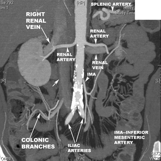
| PA view Reconstructed image | Origin and course of renal Arteries |
| PA view Reconstructed image | Origin and course of renal Arteries |
| PA view Reconstructed image | Origin and course of renal Arteries |
| CT | Note the origin of Renal arteries from the lateral aspect of Aorta |
| CT | Origin of left renal Artery |
| Branches | |
| Relationship | |
| CT | Note the relationship to Renal Veins |
| Appearance in | |
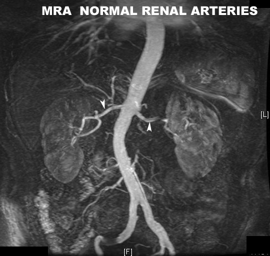
| PA view Reconstructed image MRA | Origin and course of renal Arteries |
| Course | |
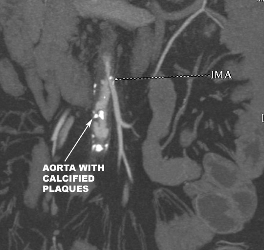
| PA view Reconstructed image | Identify Inferior Mesenteric artery |
| Lateral view Reconstructed image | Identify Inferior Mesenteric artery |
| PA view Reconstructed image | IMA |
| Branches | |
| Relationship | |
| Appearance in | |
| Drawing or CT | What organs are supplied by Inferior Mesenteric artery |
| Drawing or CT | What organs are supplied by Celiac artery |
| Course | |
| PA view Reconstructed image | Start and end of Iliac artery |
| PA view Reconstructed image | Identify branches of Iliac artery |
| CT | Distal Aorta |
| CT | Aortic Bifurcation |
| CT | Aortic Bifurcation |
| CT | Common Iliac arteries |
| Branches | |
| CT | Divides into internal and external Iliac |
| Relationship | |
| CT | Note the accompanying vein |
| CT | Note the accompanying vein |
| CT | Note the accompanying vein |
| Appearance in | |
| Internal iliac artery | |
| Course | |
| CT | Internal iliac travels posteriorly |
| Branches | |
| CT | Divides into internal and external Iliac |
| Relationship | |
| Appearance in | |
| External Iliac artery | |
| Course | |
| CT | External Iliac travels anteriorly |
| CT | External Iliac travels anteriorly |
| CT | External Iliac becomes Femoral artery |
| Branches | |
| Relationship | |
| Appearance in | |
| CT | Femoral artery and its major branch |
| Image174a | |
| Drawing or CT | Which organs does it drain |
| Course / Origin | |
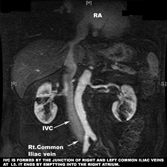
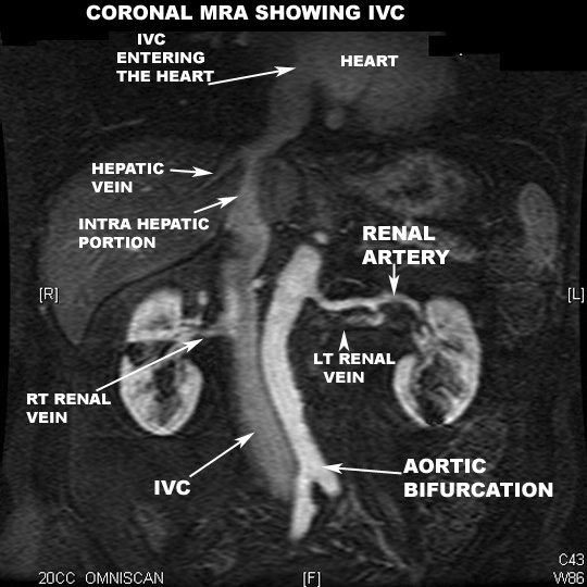
| Reconstructed image MRA | Where does it start and end |
| CT | Common Iliac veins |
| CT | Common Iliac veins coming together |
| CT | Iliac veins join to form IVC |
| CT | IVC has formed |
| Tributaries | |
| PA view Reconstructed image | Tributaries emptying into IVC |
| Reconstructed image MRA | Tributaries emptying into IVC |
| Relationship | |
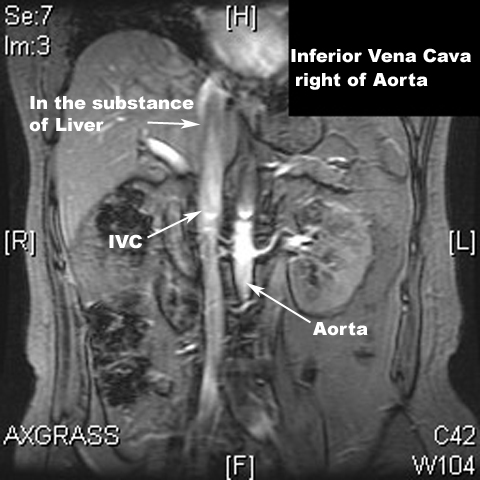
| Reconstructed image | Relationship to Aorta |
| CT | Relationship of IVC near diaphragm |
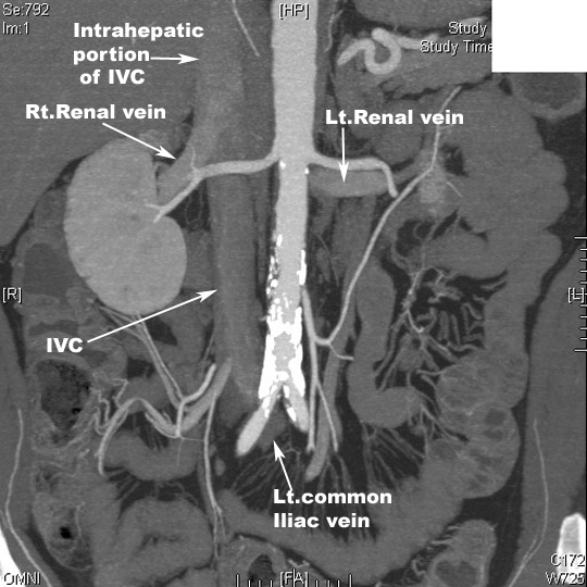
| CT | Relationship of IVC near Kidney. Renal veins entering IVC |
| Appearance in | |
| US | US IVC |
| US | US IVC |
| Color Doppler | Note the relationship of inferior vena cava to pancreas |
| Color Doppler | Hepatic veins draining into IVC |
| Iliac Veins | |
| Course / Origin | |
| CT | Common Iliac Vein |
| CT | Common Iliac Vein |
| CT | Common Iliac Vein |
| CT | Common Iliac Vein |
| CT | External Iliac vein |
| CT | External Iliac vein |
| Tributaries | |
| Relationship | |
| Appearance in | |
| Drawing or CT | Which organs does it drain |
| Course / Origin | |
| Reconstructed image | Where does it start and end |
| Reconstructed image | What are the tributaries joining Portal vein |
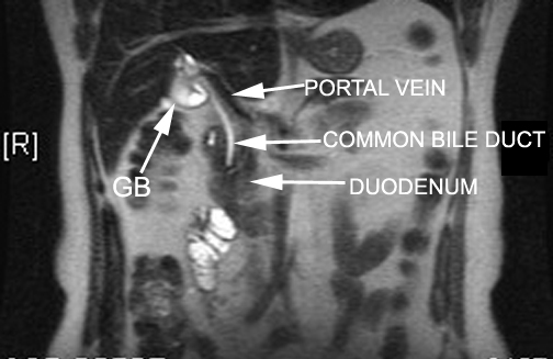
| CT | Portal vein in Porta Hepatis |
| CT | Portal vein in Porta Hepatis |
| CT | Left branch of Portal vein |
| CT | Right and left branch of Portal vein |
| CT | Portal vein |
| US | Portal Vein |
| US | Portal Vein |
| Tributaries | |
| CT | Splenic vein joining SMV to form Portal vein |
| CT | Splenic vein joining SMV to form Portal vein |
| CT | Splenic vein joining SMV to form Portal vein |
| CT | Beginning of Portal vein |
| Relationship | |
| CT | Anterior to caudate lobe |
| US | Relationship to CBD |
| CT | Note the relationship of Portal vein to IVC |
| Color Doppler | Relationship to CBD |
| Appearance in | |
| Color Doppler | Portal triad |
| Course / Origin | |
| CT | Right, middle and left hepatic veins |
| CT | Playboy bunny |
| CT | Middle and left hepatic veins |
| US | Hepatic veins draining into IVC |
| US | Hepatic veins draining into IVC |
| US | Hepatic veins draining into IVC |
| US | Hepatic veins draining into IVC |
| US | Hepatic veins d |
| Tributaries | |
| Relationship | |
| Appearance in | |
| CT | Hepatic and portal veins |
| Course / Origin | |
| Reconstructed image | Course of Renal veins |
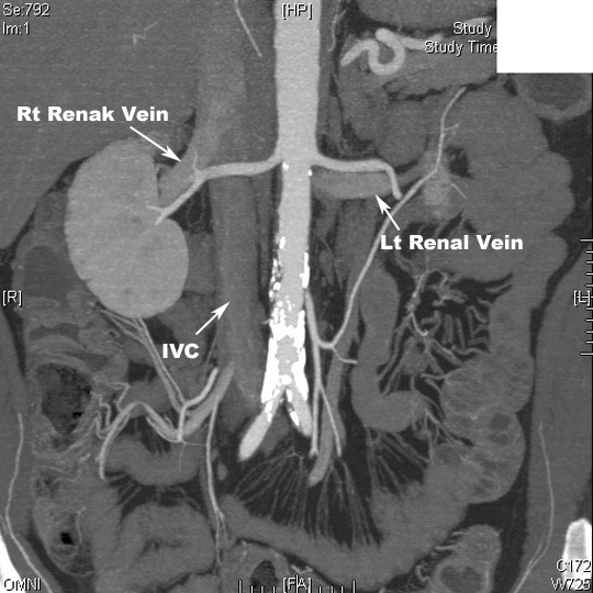
| PA view Reconstructed image | Renal veins entering IVC |
| CT | Renal veins entering IVC |
| CT | Renal veins entering IVC |
| CT | Left renal vein draining into IVC |
| Tributaries | |
| What other tributaries join Renal veins? | |
| Relationship | |
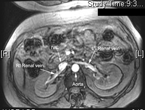
| MR | Left Renal vein crossing Aorta to join IVC |
| CT | Left renal vein crossing in front of Aorta |
| Appearance in | |
| Course / Origin | |
| CT | Splenic vein joining SMV to form Portal vein |
| CT | Splenic vein joining SMV to form Portal vein |
| CT | Origin and end of Splenic vein |
| Tributaries | |
| Relationship | |
| CT | Relationship of Splenic vein |
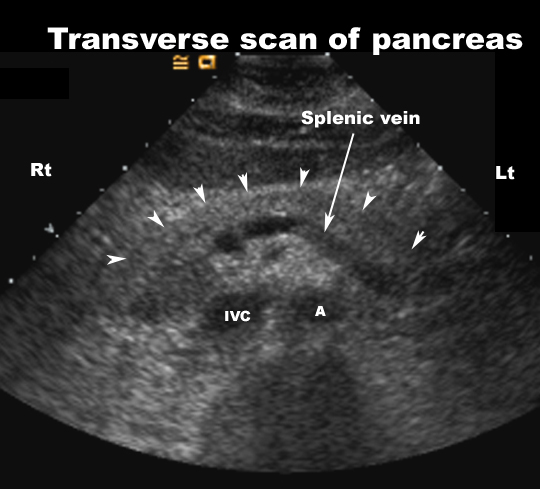
| US | Relationship to Pancreas |
| Appearance in | |
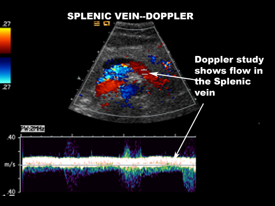
| Color Doppler | Relationship of Splenic vein |
| CT | Splenic vein |
| US | Splenic Vein |
| US | Splenic Vein |
| Salena7a | |
| Salena5a | |
| Color Doppler | Splenic vein draining into IVC |