|
This has two modules
This exercise is for students studying Anatomy. Try answering the questions first .Click on the image to see the labeled image with answers. The objective is to learn Anatomy using Radiological images.. |
|
| Location | |
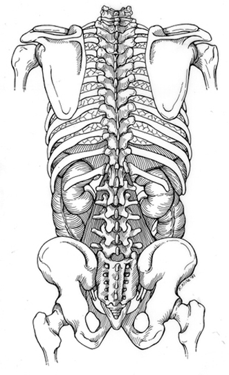
| Image 1 | Note the location of Kidneys |
| Image 2 | Identify Kidney in this Image. Located in retro peritoneum. |
| Image 3 | Which Kidney is higher in this IVP? |
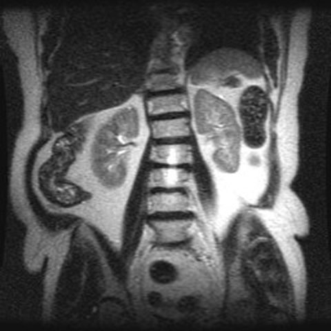
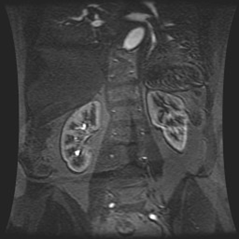
| Image 4 | Appreciate the higher position of the left Kidney in this MR? |
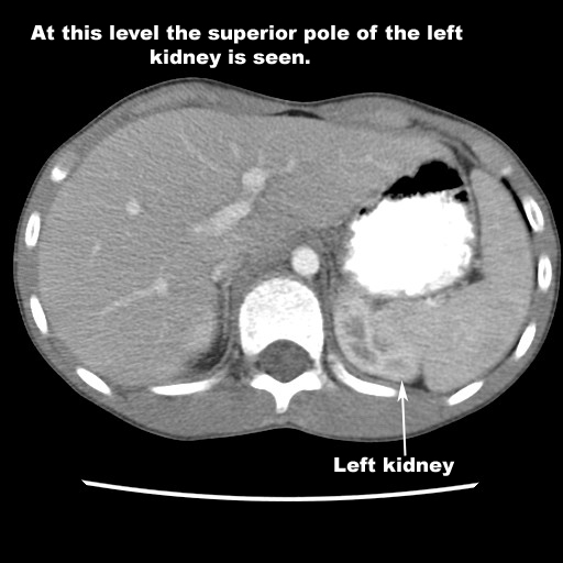
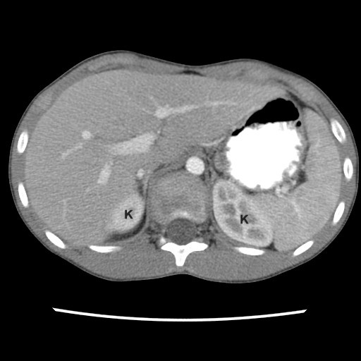
| Image 5 | In CT, left kidney will appear first as it is higher in position |
| Image 6 | Right Kidney will appear in inferior sections. |
| Shape | |
| Image 1 | Autopsy specimen showing the shape of Kidney |
| Image 2 | Appreciate the shape of Kidney in this IVP |
| Image 3 | Appreciate the shape of Kidney in this MR |
| Size | |
| Image 1 | Best imaging procedure to measure the size of Kidneys |
| Axis | |
| Image 1 | Draw the axis of Kidney in this IVP |
| Structure | |
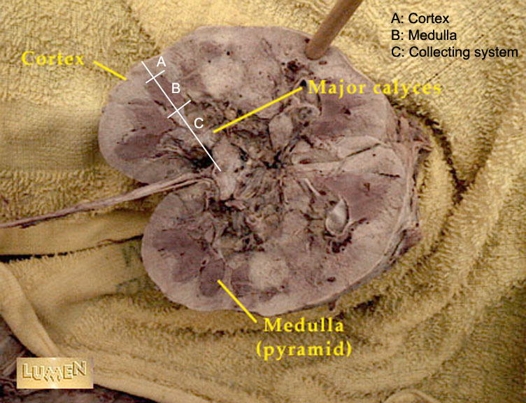
| Image 1 | Autopsy specimen showing Cortex and Medulla |
| Image 2 | Identify Cortex and Medulla in this CT |
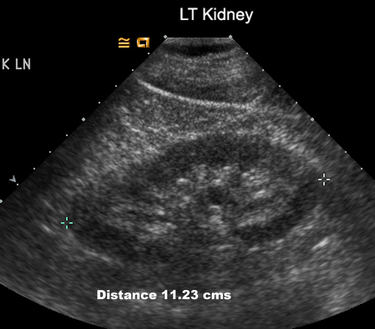
| Image 3 | Identify Cortex and Medulla in US |
| Image 4 | MR Showing outer cortex and inner dark Medulla |
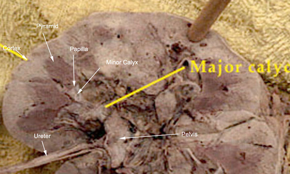
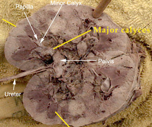
| Image 5 | Autopsy specimen showing Pyramid, Papilla and Calyx |
| Image 6 | Identify Pyramids in US |
| Image 7 | Identify Pyramids in US |
| Relationship | |
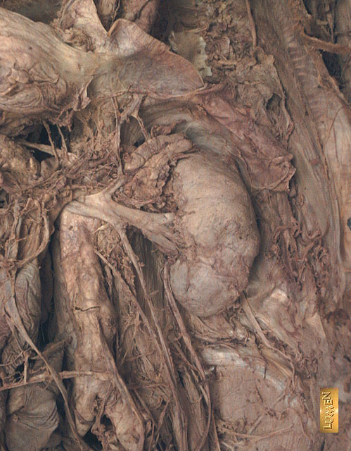
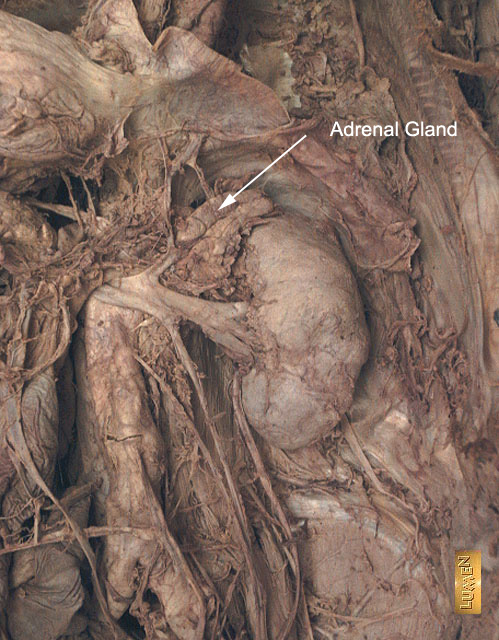
| Image 1 | Autopsy specimen showing location of the Adrenal glands |
| Image 2 | Identify Adrenals in CT. They appear in sections above Kidney. |
| Image 3 | Note the relationship of Kidney to Colon, Duodenum, Liver, Spleen, Adrenal, Pancreas in CT |
| Image 4 | Note the relationship of Kidney to Liver, Spleen, Aorta, Stomach in CT |
| Peri-nephric | |
| Image 1 | Identify peri-nephric Fat in CT |
| Image 2 | Identify peri-nephric Fat in this MR |
| Renal Collecting system | |
| Kidney | |
| Image 1 | Autopsy specimen of Kidney showing the Renal collecting system |
| Image 2 | Identify Fornix, Calyx, Pelvis, Ureter in IVP |
| Image 3 | Identify Collecting system in MR |
| Ureter | |
| Image 1 | Identify Pelvis, Ureters, Bladder in IVP |
| Image 2 | Identify Ureters, Psoas in CT |
| Image 3 | Identify the Ureters |
| Bladder | |
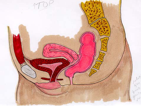
| Image 1 | Showing bladder uterus relationship |
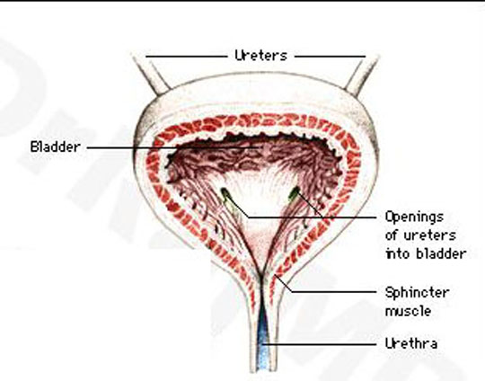
| Image 2 | Note Ureters entering Bladder |
| Image 3 | Note Ureters entering Bladder |
| Image 4 | Identify Bladder, Urine jet in US |
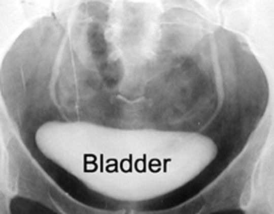
| Image 5 | Identify Bladder and Urethra in Cystourethrogram |
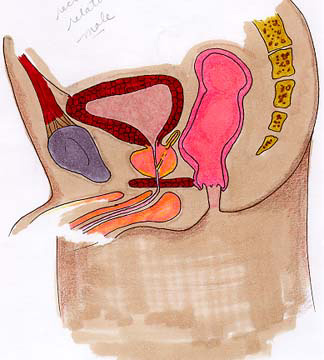
| Image 6 | Identify Rectum, Bladder, Uterus in MR |
| Image 7 | Identify the structure between Rectum and Bladder |
| Prostate | |
| Image 1 | Identify Rectum, Prostate in CT |
| Urethra | |
| Image 1 |
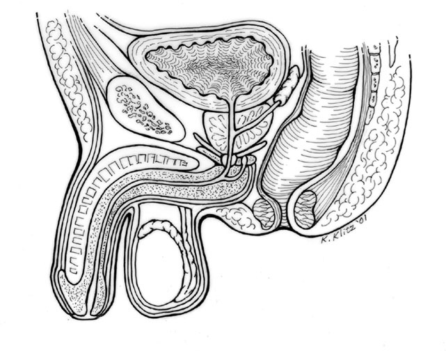
Male urethra: |
| Image 2 | Urethra is encircled by prostate, as it leaves bladder |
| Image 3 | Identify various portions of male Urethra in this voiding Cystourethrogram |
| Image 4 | Note the short female Urethra in this voiding Cystourethrogram |
| Renal vasculature | |
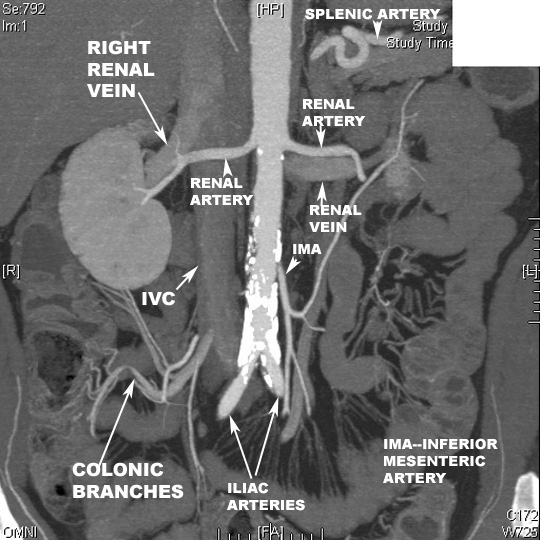
| Image 1 | Identify the branches of Abdominal Aorta in this reconstructed CT Angiogram image |
| Image 2 | Note the origin and course of Renal arteries and Renal veins |
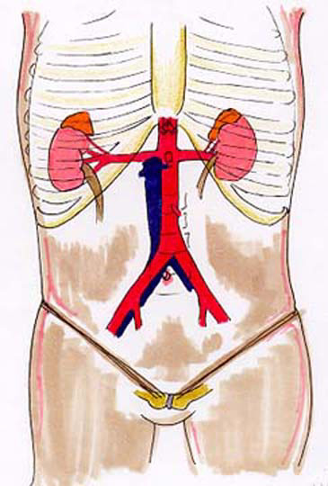
| Image 3 |
Diagram of Renal arteries |
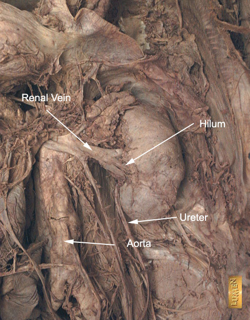
| Image 4 | Autopsy specimen showing the hilum of Kidney with Renal vessels |
| Image 5 | Identify Renal arteries in this reconstructed MR Angiogram image |
| Image 6 | Identify Renal artery, Aorta, IVC in MR |
| Image 7 | Identify Renal veins, IVC, Aorta in MR |
| Image 8 | Identify Renal artery, Aorta, Vena cava in CT angiogram |
| Image 9 | Identify Renal vein, IVC in CT angiogram |
| Image 10 | Identify Renal artery and vein in Color Doppler Ultrasound |
| Image 11 | Identify Arterial, Venous, Collecting system in Color Doppler Ultrasound |
| Scrotum | |
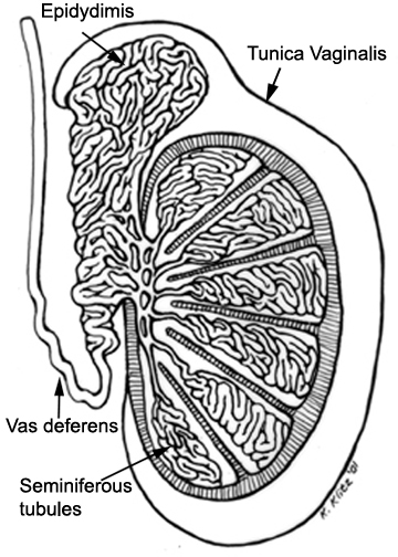
| Image 1 |
Drawing showing Testis Epididymis relationship |
| Image 2 | Drawing showing Testis Epididymis relationship |
| Image 3 | Identify Testis, Epididymis in US |
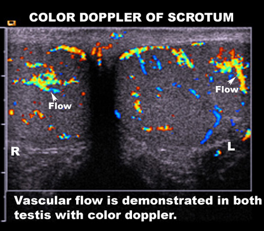
| Image 4 | Color Doppler showing blood flow to Testis |