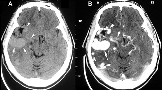
Arterial Venous Malformation
- A: Non-contrast CT shows a lobular mass in the right hemisphere. The arrowheads in A point to nonenhanced vessels.
- B: Post-contrast demonstrates intense enhancement of the mass with multiple prominent vascular channels (arrowheads) - large AVM.