Pancreatitis
Pathology
- Pancreas is edematous and enlarged.
- Pancreas can show extensive necrosis, acute inflammation, suppuration and/or hemorrhage.
- There can be extensive peripancreatic inflammation.
- Fluid can accumulate in lesser sac and pleural space and paracolic gutters.
- Neutrophils infiltrate the edge of the necrotic areas and extend into the adjacent lobules of fat and produce fat necrosis.
- Calcification can be seen in chronic pancreatitis.
Potential complications
- Abscess/pseudocyst
- As liquefaction of necrotic pancreatic tissue progresses, it will gradually take on the appearance of localized fluid collection - pseudocyst
- This may be in the region of the pancreas or extend beyond the pancreatic region
- Pancreatic rupture/hemorrhage
- Obstructive jaundice
- Pulmonary complications in severely ill patients - ARDS
- GI obstruction
- Acute renal failure
|
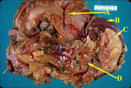 |
An autopsy specimen consisting of the stomach (A), spleen (B), pancreas (D), and adjacent fat (C) reveals acute inflammation. The pancreas is swollen and hyperemic. Focal areas of green necrosis are present. Small foci of bright yellow, fat necrosis are present. The stomach is folded back so as to reveal its posterior wall and the pancreas. |
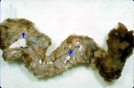 |
Chronic pancreatitis: The pancreas is shrunken and fibrotic. The main duct is dilated and filled with calcified secretions (small arrows). |