|
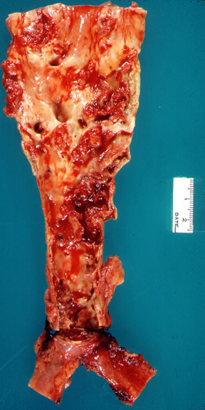
The abdominal aorta was opened along the posterior wall revealing severe atherosclerosis. The anatomical features are obscured by ulcerated plaques and superimposed blood clots. |
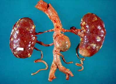 |
A medium size aneurysm arises from the aorta between the renal and inferior mesenteric arteries. |
What are the anticipated imaging findings of abdominal aortic aneurysm?Widened aortic lumen
Additional findings
|
|
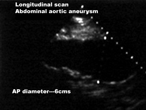 |
Ultrasound showing findings of Abdominal Aortic Aneurysm
|
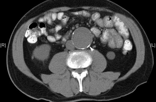 |
CT scan from another case with Abdominal Aortic AneurysmNote wall calcification of the aneurysm of aorta.
|
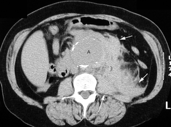 |
Ruptured Abdominal Aortic AneurysmCT without IV contrast shows abdominal aortic aneurysm (A) with high density blood (arrows) indicating rupture.
|