|
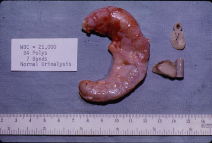
The appendix is markedly swollen. The serosa is hyperemic and covered by a fibrinous exudate. Compare the inflammed appendix to the adjacent segments of a normal appendix. |
What are the anticipated findings of appendicitis in imaging procedures?Imaging Findings:
|
|
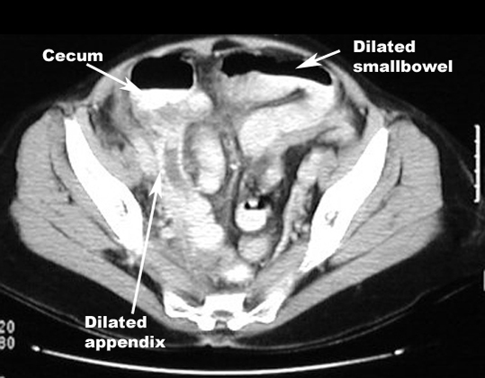 |
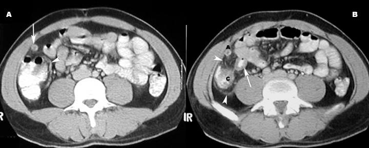
Acute AppendicitisCT scan showing
|
 |
CT scan showing Appendicolith
|
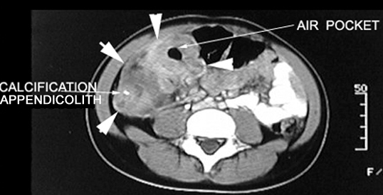 |
CT scan showing findings of appendiceal abscess.Arrows point to the inflammatory mass in the right lower quadrant with an air pocket, indicating an abscess. Mass demonstrates contrast enhancement. |