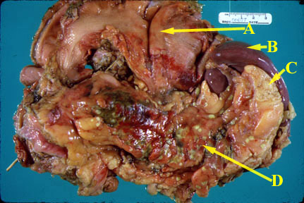
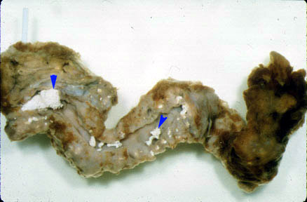
Pathology
- Pancreas is edematous and enlarged.
- There can be extensive peripancreatic inflammation.
- Pancreas can show acute inflammation, suppuration and/or hemorrhage and extensive necrosis.
- Neutrophils infiltrate the edge of the necrotic areas and extend into the adjacent lobules of fat and produce fat necrosis.
- Fluid can accumulate in lesser sac and pleural space and paracolic gutters.
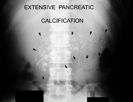
- Calcification can be seen in chronic pancreatitis.
Potential acute complications
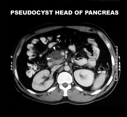
- Abscess/pseudocyst
- As liquefaction of necrotic pancreatic tissue progresses, it will gradually take on the appearance of localized fluid collection - pseudocyst
- This may be in the region of the pancreas or extend beyond the pancreatic region
- Pancreatic rupture/hemorrhage
- Obstructive jaundice
- Pulmonary complications in severely ill patients - ARDS
- GI obstruction
- Acute renal failure
What are the anticipated imaging findings of acute pancreatitis?
- Abdominal x-ray is not diagnostic, but may
show:
- Calcification in the pancreas
- Mass from a pseudocyst
- Sentinel loop: Dilatation of duodenum
- Colon cut off: Dilated colon to the mid-transverse colon. No air seen beyond splenic flexure. this is due to extension of inflammation along mesocolon.
- Diffuse ileus ( small bowel dilatation) most commonest
- Pleural effusion
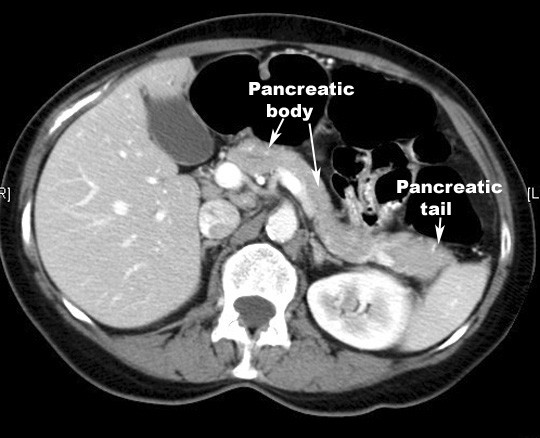
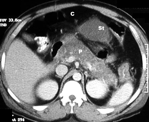
- Contrast-enhanced CT of the pancreas is
diagnostic and can show:
- Enlargement of pancreas due to edema
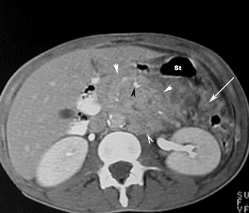
- Peripancreatic inflammation: linear strands in the peripacreatic fat
- Phlegmon
- Hemorrhagic: Enlarged pancreas with increased density due to hemorrhage
- Necrosis: On contrast enhanced phases the necrotic pancreatic parenchyma will show decreased or no enhancement when compared with normally enhancing viable tissue
- Fluid in the paracolic gutter
- Fluid collections: A simple peripancreatic fluid collection will not have a well-defined capsule
- Pseudocysts: As liquefaction of necrotic pancreatic tissue progresses it will gradually take on the appearance of localized fluid collection...pseudocyst
- Abscesses: Diffusely enlarged pancreas with air pockets