Pancreatitis

Pathology
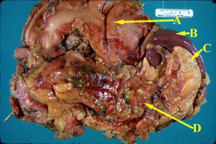
  • Pancreas is edematous and enlarged.
  • There can be extensive peripancreatic inflammation.
  • Pancreas can show acute inflammation, suppuration and/or hemorrhage and extensive necrosis.
  • Neutrophils infiltrate the edge of the necrotic areas and extend into the adjacent lobules of fat and produce fat necrosis.
  • Fluid can accumulate in lesser sac and pleural space and paracolic gutters.
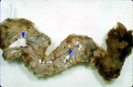
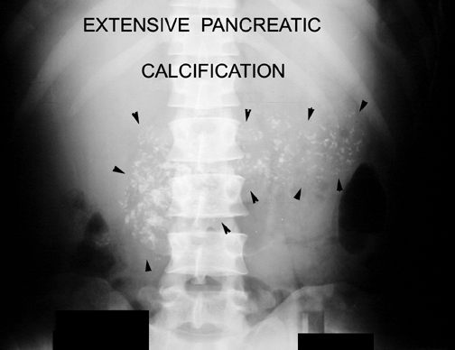
  • Calcification can be seen in chronic pancreatitis.
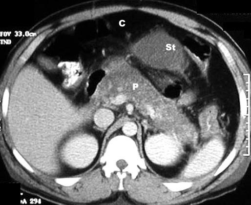
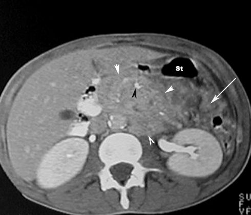
Potential acute complications
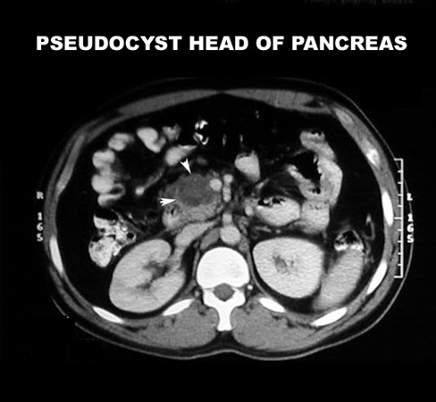
  • Abscess/pseudocyst
    • As liquefaction of necrotic pancreatic tissue progresses, it will gradually take on the appearance of localized fluid collection - pseudocyst
    • This may be in the region of the pancreas or extend beyond the pancreatic region
  • Pancreatic rupture/hemorrhage
  • Obstructive jaundice
  • Pulmonary complications in severely ill patients - ARDS
  • GI obstruction
  • Acute renal failure 
An autopsy specimen consisting of the stomach (A), spleen (B), pancreas (D), and adjacent fat (C) reveals acute inflammation. The pancreas is swollen and hyperemic. Focal areas of green necrosis are present. Small foci of bright yellow, fat necrosis are present. The stomach is folded back so as to reveal its posterior wall and the pancreas.
What are the anticipated imaging findings of acute pancreatitis?
  • Abdominal x-ray is not diagnostic, but may show:
    • Calcification in the pancreas
    • Mass from a pseudocyst
    • Sentinel loop: Dilatation of duodenum
    • Colon cut off: Dilated colon to the mid-transeverse cpon. No air seen beyond splenic flexure. this is due to extension of iflammation along mesocolon.
    • Diffuse ileus ( small bowel dilatation) most commonest
    • Pleural effusion
  • Contrast-enhanced CT of the pancreas is diagnostic and can show:
    • Enlargement of pancreas due to edema
    • Peripancreatic inflammation: linear strands in the peripacreatic fat
    • Phlegmon
    • Hemorrhagic: Enlarged pancreas with increased density due to hemorrhage
    • Necrosis: On contrast enhanced phases the necrotic pancreatic parenchyma will show decreased or no enhancement when compared with normally enhancing viable tissue
    • Fluid in the paracolic gutter
    • Fluid collections: A simple peripancreatic fluid collection will not have a well-defined capsule
    • Pseudocysts: As liquifaction of necrotic pancreatic tissue progresses it will gradually take on the appearance of localized fluid collection...pseudocyst
    • Abscesses: Diffusely enlarged pancreas with air pockets


Some imaging findings of acute pancreatitis:

CT scan in Acute Pancreatitis

Post Contrast CT findings reveal diffusely enlarged pancreas with low density from edema.

C:  Colon
St: Stomach
P:  Pancreas

CT scan in Acute Pancreatitis
Phlegmon / Inflammatory mass

  • White arrowheads: Phlegmon
  • Black arrowhead:   Pancreatic calcification
  • Large Arrow: Peripancreatic fascial infiltration

  St: Stomach

Potential chronic complications of pancreatitis:
  • Malabsorption
  • Pseudocyst
What are the anticpated imaging findings of chronic pancreatitis?
  • Calcifications in the pancreas
  • Pseudocysts


Pathology Chronic pancreatitis:

The pancreas is shrunken and fibrotic. The main duct is dilated and filled with calcified secretions (small arrows).

Some imaging findings of Chronic pancreatitis:

Plain x-ray abdomen in a patient with Chronic Pancreatitis

Arrowheads point to extensive pancreatic calcification.

CT scan in a patient with chronic pancreatitis and pseudocyst.