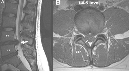
- A & B: Sagittal and axial MR images show a large herniation of intervertebral disc at L4-L5 with compressed thecal sac and nerve roots (black arrows).
- There is also a second herniation at L5-S1 seen on the sagittal image.
CT or MRI are typically ordered for patients with severe or prolonged pain, focal neuro deficits or history of cancer or febrile illness.
22 y/o patient complains of one week of back pain. The pain started the day after he helped his friend move to a different apartment. It does not wake him from sleep, and is alleviated with NSAIDS. The pain is slowly improving, and he is able to go to work without significant discomfort.
A 49 y/o female reports three weeks of lower back pain. The pain is not associated with any specific incident. The pain radiates into her left buttock and posterior thigh. There is mild weakness of left foot dorsiflexion and toe extension. The patient has a few fasciculations is her left dorsal foot. Pinprick sensation is decreased over his left dorsal foot. There is no Babinski sign. Her pain can be reproduced with passive flexion of the hip.
|
|
Herniated Lumbar Disc
|
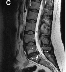
C: Another case showing a large herniated disc at L5-S1 with compression of thecal sac and nerve roots (black arrows). |
 |
 |
 |
 |
| Sagittal T 1 wtd. MRI of the lumbar spine | Post contrast (C+) sagittal T 1 wtd. MRI | Sagittal T 2 wtd. MRI | Axial T 1 wtd. MRI (C+) |
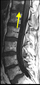
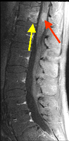
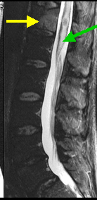
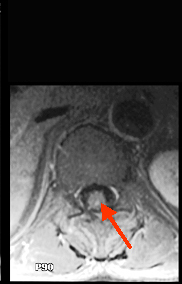
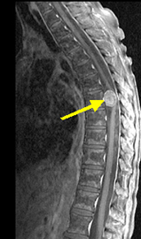
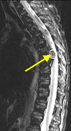
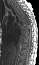
Bony Metastasis and metastasis to the Spinal CordFindings: : Bony metastasis (yellow arrow in A, B, C) is seen involving the T 12 vertebral body. Intramedullary location of metastasis within the distal thoracic cord, is verified on post contrast sagittal image (red arrow in B) and axial image (red arrow in D). Edema (green arrow in C) within the thoracic cord is best shown on T 2 wtd. image C. |
|||
40 y/o male has 1 day of spastic weakness in his legs, and one episode of incontinence. His knee and ankle deep tendon reflexes are increased (3+) bilaterally, but 2+ elsewhere. The upper limbs are normal. Pinprick and temperature sensation are decreased below mid-torso.
 |
 |
 |
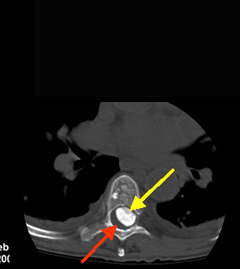 |
| Post contrast (C+) sagittal T1 wtd. MRI | Sagittal T2 wtd. MRI of thoracic spine | Precontrast sagittal T1 wtd. MRI | Axial CT image of the thoracic spine |
Classic example of calcified intradural meningiomaFindings: : An intradural enhancing meningioma (arrow in A), the ventrally located tumor has produced cord compression and with displacement of the thoracic cord (red arrow in D) to the right side. Calcified nature of the tumor is identified on sagittal T 2 wtd. image as an area of dark signal intensity (yellow arrow in B) and confirmed by CT imaging (yellow arrow in D) as an area of high attenuation density. |
|||
Case 5:
82y/o thin female with back pain for several months. Her back pain began after a minor fall that she had several months ago. She stated that she just fell into a sitting position on her couch. Her neurological exam is normal.
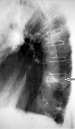 |
OsteoporosisCompression fracture of spine
|