Hydrocephalus
Hydrocephalus, or ventricular enlargement, may involve some or all of the ventricles, depending on whether there is a specific site of obstruction to CSF flow. The ventricles appear enlarged. Significant loss of brain tissue can also lead to enlarged ventricles.
Describe the CSF pathway.
- Cerebrospinal fluid is produced in the lateral, thrid and fourth ventricles by the choroid plexus.
- CSF travels from the lateral ventricles through the interventricular foramina to the third ventricle, then through the cerebral aqueduct to the fourth ventricle.
- CSF then leaves the ventricular system at the fourth ventricle through the midline foramina of Magendie and the paired lateral foramina of Luschka.
- The CSF enters the subaracnoid spaces where it circulates to bathe the surface of the brain and spinal cord.
- The CSF leaves the subarachnoid space and enters the venous sinuses by unidirectional valves called arachnoid villi.
What is hydrocephalus?
- Hydrocephalus is defined as an increase in the volume of CSF within the ventricular system.
What are the types of hydrocephalus?
- Obstructive hydrocephalus
- Communicating hydrocephalus
What are the characteristics of obstructive hydrocephalus ?
- The block of CSF flow occurs within the ventricular system or at the foramina that exit the fourth ventricle and the ventricular system enlarges proximal to the obstruction.
- An example of this is aqueductal stenosis, where there is enlargement of the lateral and third, but not the fourth ventricles.
What are the consequences of obstructive hydrocephalus in children?
- Obstructive hydrocephalus results in increased intracranial pressure, which results in increased head circumference by widening of the cranial sutures, in an attempt to decrease the pressure.
- The increased pressure also results in tense fontanelles.
- Signs and symptoms of hydrocephalus include emesis (especially in the morning), decline in cognitive ability, headaches, papilledema, ataxia, and defect in upward gaze.
What are the characteristics of communicating hydrocephalus?
- Although there is no obstruction of the CSF pathway, CSF reabsorption is impaired in communicating hydrocephalus, at the basal cisterns or at the surfaces of the cerebral hemispheres.
- An example is scarring at the arachnoid villi from previous bleeding.
- Clinical features of communicating hydrocephalus are like those of obstructive hydrocephalus.
Case 1:
Hydrocephalus Obstructive
Clinical:
F-54 with delayed milestones, S/P occipital decompression of Chiari malformation, with progressive gait ataxia, adduction paresis of eyes, nystagmus and dementia
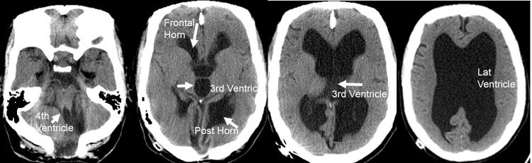
CT scans without contrast show enlargement of lateral and third ventricles, but not the fourth ventricle, due to aqueductal stenosis. The posterior fossa is distorted due to a congenital Chiari malformation and previous surgery.