|
N- |
|
|
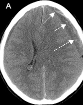

Diagnosis: Chronic Subdural Hematoma
55 year-old patient with chronic myelogenous leukemia with low platelet count.
- A: Left frontal chronic subdural hematoma (arrows) seen as an area of low-density with crescentic inner margin, compressing the adjacent brain.
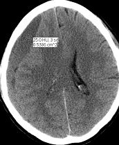
- B: Left frontal subdural hematoma was completely evacuated using burr holes in the skull, but the right chronic subdural hematoma has increased in size in the follow-up CT done 19 days later (arrows) which was also subsequently evacuated.