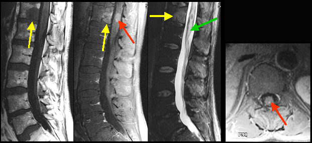
Bony Metastasis and metastasis to the Spinal Cord
Findings: : Bony metastasis (yellow arrow in A, B, C) is seen involving the T 12 vertebral body. Intramedullary location of metastasis within the distal thoracic cord, is verified on post contrast sagittal image (red arrow in B) and axial image (red arrow in D). Edema (green arrow in C) within the thoracic cord is best shown on T 2 wtd. image C.