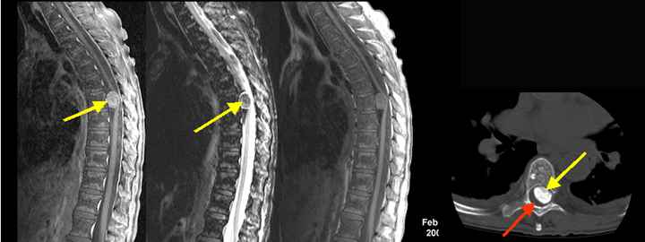
Classic example of calcified intradural meningioma
Findings: : An intradural enhancing meningioma (arrow in A), the ventrally located tumor has produced cord compression and with displacement of the thoracic cord (red arrow in D) to the right side. Calcified nature of the tumor is identified on sagittal T 2 wtd. image as an area of dark signal intensity (yellow arrow in B) and confirmed by CT imaging (yellow arrow in D) as an area of high attenuation density.