Ovarian Tumors 

What is the clinical setting when you will consider an ovarian mass?
What are the common ovarian masses?
What are the useful imaging modalities in evaluating an ovarian mass?
What is the utility of each procedure?

The radiographic evaluation of the patient depends on the age and clinical suspicion.  

What are the Imaging findings of ovarian cyst?
What are the radiological findings of a malignant ovarian mass?

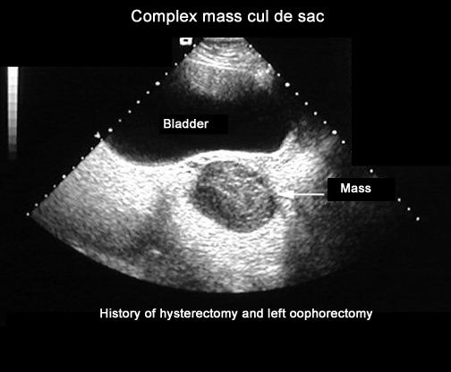
Any solid component on the ultrasound is a possible feature of malignancy. 


Complex cystic and solid mass
A thick wall can be seen, but some benign cysts also have a thick wall. 

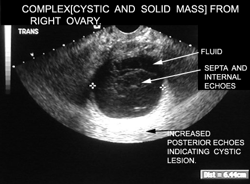
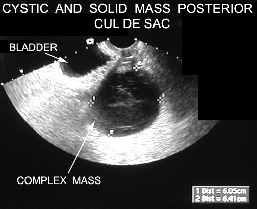
The other characteristics that are associated with malignancy are septations. 

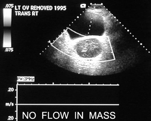
Doppler also improves the specificity of ultrasound.

Malignant masses are more likely to have detectable flow and flow that is centrally located. 

No flow in mass.
Case 1:
Patient is a 37 yr/o G3P2012 who comes to the ER complaining of severe abdominal pain.  Patient has had a similar pain in the past.  Patient has regular periods, which come every 28-30 days. Her LMP was 2 weeks ago.  She is afebrile.  On physical exam, she has right lower quadrant tenderness, with no rebound or guarding. Patient has no cervical discharge, no cervical motion tenderness, but does have right adenxal tenderness. Resident ordered CT.
Case 2:
Patient is a 48 yr/o G0P0 who presents to her gynecologist with an increase in abdominal girth.  She has had no pain, but does complain of increase in urinary frequency.  On further questioning, she states that she has had increasing constipation over the past several weeks.  On pelvic exam, patient is noted to have large right sided adenxal mass.  US which shows the large ovarian mass.
This patient had a lymphoma. CT of abdomen and pelvis which shows the mets