Ovarian Tumors
by
Molly Jonna, MS4 and Jennifer Lim-Dunham, M.D.
What are the common ovarian masses?
- Ovarian cyst
- Germ cell tumors including Dermoid cyst and Teratoma
- Other ovarian cancer
What are the useful imaging modalities in evaluating an ovarian mass?
- Pelvic Ultrasound
- MRI
- Pelvic CT
What is the utility of each procedure?
The radiographic evaluation of the patient depends on the age and clinical suspicion.
- Ultrasound:
- It is the primary way to evaluate pelvic masses.
- Transvaginal US and transabdominal US are complementary, with transvaginal US giving the most accurate evaluation.
- Doppler US, which looks for blood flow into the ovary, is a useful adjunct.
- MRI:
- MRI can characterize pelvic masses with respect to solid and cystic components, and determine their organ origin. Distinguishes benign from malignant ovarian masses with 91% accuracy.
- No ionizing radiation
- CT:
- Not indicated for the primary evaluation of adnexal masses, because it is difficult to characterize ovarian masses
- However, in cases of known or suspected ovarian cancer, CT is very good technique in evaluating the extent of metastatic disease
Appropriateness Criteria:
The American College of Radiology has developed Appropriateness Criteria which are evidence-based guidelines that assist physicians in making the most appropriate imaging decisions for a wide variety of clinical conditions.
Appropriateness Criteria for clinically suspected adnexal mass states that transvaginal and transabdominal ultrasonography with Doppler is the test of choice. MRI is useful for further characterization of adnexal masses, while CT is rarely indicated for primary evaluation. Links to the criteria can be found below.
Link to women’s imaging recommendations:
http://www.acr.org/Quality-Safety/Appropriateness-Criteria/Diagnostic/Womens-Imaging
Link to adnexal mass imaging recommendations:
http://www.acr.org/~/media/ACR/Documents/AppCriteria/Diagnostic/ClinicallySuspectedAdnexalMass.pdf
What are the Imaging findings of ovarian cyst?
- On ultrasound, simple cysts are characterized by anechoic (black) fluid filling the cyst cavity and thin walls.
- If an ovarian cyst has recently ruptured, one will see fluid in the pelvis.
- If there are echoes within the cyst, it may be from hemorrhage.
What are the radiologic findings of a malignant ovarian mass?
- Any solid component on the ultrasound is a possible feature of malignancy
- Complex cystic and solid mass
- A thick wall can be seen, but some benign cysts also have a thick wall
- Solid components of the mass are more likely to have centrally located detectable flow on color Doppler ultrasound
What are ovarian germ cell tumors?
- Teratomas are germ cell tumors commonly composed of multiple cell types derived from one or more of 3 germ layers. They are almost always benign They occur most frequently during the reproductive years and are often discovered incidentally.
- Complications of teratomas are torsion, rupture, infection, hemolytic anemia, and malignant degeneration.
- A dermoid is a mature cystic teratoma composed of developmentally mature skin with hair follicles and sweatglands, sometimes clumps of long hair, and often pockets of sebum, blood, fat,bone, nails, teeth, eyes, cartilage, and thyroid tissue. Because it contains mature tissue, a dermoid cyst is almost always benign.
What are the imaging findings in dermoid cysts and ovarian teratomas?
- Predominately cystic mass that may be unilocular or multilocular.
- May contain calcifications with acoustic shadows as a result.
- May contain hyper echoic lines (dermoid mesh) representing hair floating in the fluid
- May contain a fat-fluid level, i.e., fat floating in aqueous fluid
- May contain echogenic solid nodule (dermoid plug), which contains calcific, dental, adipose, hair and/or sebaceous components.
- May demonstrate the “tip-of-the iceberg” sign, which refers to prominent acoustic shadows produced by highly echogenic hair and sebum.
- CT and MRI imaging shows calcifications with foci of fat.
What is the clinical setting when you will consider an ovarian mass?
- You would consider an ovarian mass in any woman who comes in complaining of pressure symptoms.
- These symptoms include urinary frequency, pelvic discomfort, and constipation.
- Abdominal swelling, along with fatigue and abdominal pain are the most common symptoms.
- Irregular vaginal bleeding can accompany any ovarian neoplasm.
- Some of the ovarian tumors (Sertoli-Leydig and Granulosa) secrete androgens and estrogens, respectively.
- With the Sertoli-Leydig tumors a woman may present not only with an abdominal mass, but also with virilization.
Take Home Points:
- Pelvic ultrasound, especially transvaginal, with color Doppler is the modality of choice for evaluating adnexal masses
- A simply ovarian cyst appears as an anchoic round structure with thin walls on ultrasound
- A complex cystic and solid mass with scentrally located detectable Doppler flow is more likely to be malignant
- On ultrasound of a teratoma or dermoid cyst, one may see a dermoid mesh, a dermoid plug, and shadowing from calcifications.
Dermoid Imaging
 |
Dermoid
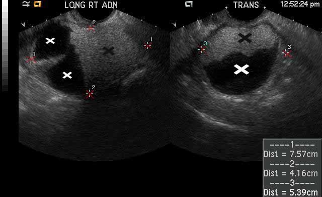
Image 1
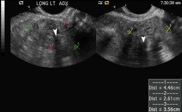
The large right adnexal dermoid cyst depicted in this image between the red calipers has both fat and fluid components, which is a classic finding. The white x’s demarcate the hypochoic, or dark, fluid components of the cyst and the black x’s demarcate the echogenic, or white, fat components of the cyst. |
 |
Dermoid
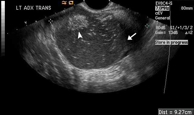
Image 2
The left adnexal dermoid cyst has both fat and fluid components as well. The white arrowhead points towards an echogenic fat component. The white arrow points toward a fluid-fluid level that separates simple from complex fluid within the cyst. The echogenic, or white, dots and dashes that you see within the cyst represent hair within the cyst. This finding is known as a “dermoid mesh. “ |
Teratoma Imaging
 |
Ovarian Teratoma
Image 1
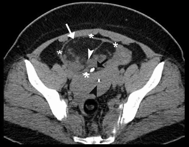
The CT image shows a large ovarian teratoma between the white calipers. The white arrow points towards a hypodense fat component and the white arrowhead points towards an isodense solid component. The black arrow is pointing towards a hyperdense calcification, which is a classic teratoma finding. The black arrowhead shows a metallic,hyperdense IUD located within the patient’s uterus. |
 |
Ovarian Teratoma
Image 2
The US image shows a teratoma which is outlined by the calipers. The white arrowheads point out hyperechoic calcification with posterior shadowing within the teratoma. The calibers are outlining the measurements of the tumor. |
Ovarian Cyst Imaging |
 |
Simple ovarian cyst
Ultrasound
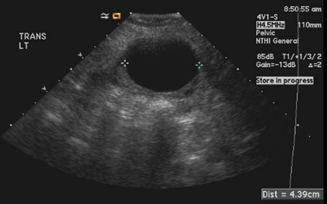
Calipers demarcate a 4.3 cm fluid-filled cyst arising from the left ovary which demonstrates the features of a simple cyst: anechoic, or completely black echotexture; thin, imperceptible wall; and posterior acoustic shadowing in which the tissues posterior to the cyst are more echogenic, or whiter, than the surrounding tissue.
|
|
Ovarian Cyst
Case 1
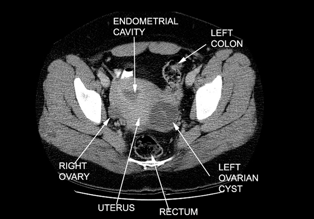
Patient is a 37 yr/o G3P2012 who comes to the ER complaining of severe abdominal pain. Patient has had a similar pain in the past. Patient has regular periods, which come every 28-30 days. Her LMP was 2 weeks ago. She is afebrile. On physical exam, she has right lower quadrant tenderness, with no rebound or guarding. Patient has no cervical discharge, no cervical motion tenderness, but does have right adenxal tenderness.
CT pelvis . Left ovarian cyst. |
Helpful links with Additional Imaging:
- http://emedicine.medscape.com/article/404450-overview#a22
- http://www.med-ed.virginia.edu/courses/rad/edus/pelvic1.html
- http://radiopaedia.org/articles/mature-cystic-ovarian-teratoma - Imaging is found on the right side of the article
- http://pubs.rsna.org/doi/figure/10.1148/radiographics.21.2.g01mr09475
References:
- Gershenson, D. M. (2013). Ovarian germ cell neoplasms: Pathology, clinical manifestations, and diagnosis. In S. Falk, B. Goff, A. Pappo & R. Garcia (Eds.), UpToDate. Retrieved from http://www.uptodate.com/contents/ovarian-germ-cell-neoplasms-pathology-clinical-manifestations-and-diagnosis?source=search_result&search=Teratoma&selectedTitle=1~88
- Outwater, E. K., Siegelman, E. S., & Hunt, J. L. (2001). Ovarian teratomas: Tumor types and imaging characteristics. RadioGraphics, 21(2), Retrieved from http://pubs.rsna.org/doi/full/10.1148/radiographics.21.2.g01mr09475
12.17.13