Hepatic Mass
What are the common liver masses?
- Malignant liver tumors
- Metastatic tumors
- Hepatocellular carcinoma
- Cholangiocarcinoma
- Benign focal liver lesions
- Cysts
- congenital
- parasitic - echinococcal
- Cavernous hemangiomas
- Focal nodular hyperplasia
- Hepatic adenomas
- Abscesses
- Cysts
What are the useful imaging modalities to investigate liver masses?
- CT
- Ultrasound
- MRI
Utility of each procedure - Indicate when you would select each procedure.
- CT scan
- Is the best imaging modality to evaluate liver masses. It can identify other intra-abdominal masses and vascular involvement, i.e., thrombosis, etc.
- CT angiography shows early enhancement of tumor in arterial phase.
- Ultrasound
- Is most useful intra-operatively, during which the transducer is placed directly upon the liver surface to recognize liver lesions.
- Transabdominal ultrasonography is inferior in sensitivity for liver masses to CT or MRI.
- Doppler is helpful to demonstrate vascularity of lesions.
- Carcinoma can be recognized as discrete nodules.
- These typically appear as slightly hyperechoic and hypoechoic nodules.
- MRI
- MRI is useful for delineating vascular involvement and identifying additional intra-abdominal lesions.
Pathology of hepatoma (hepatocellular carcinoma):
- Hepatoma can present as single or multiple masses or diffuse involvement.
- It has a tendency to invade portal and hepatic veins.
Image Atlas of Hepatic Masses
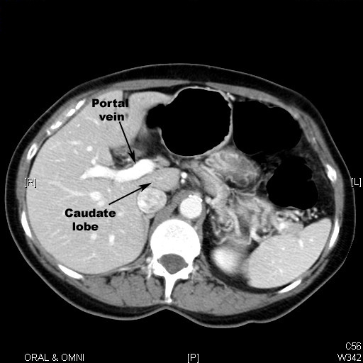
Normal Liver
- It is divided into four lobes of unequal size and shape.
- It is important to understand the complex blood flow (hepatic and portal systems) through the liver.
- Parenchyma (reticuloendothelial cells) enhances with contrast uniformly with portal vein and hepatic artery branches seen through it.
- In the superior slices we can see hepatic veins draining into inferior vena cava.
- It is of same density as spleen.
- Normal biliary ducts are not seen. They are seen only when they are dilated.
What are the imaging findings of hepatoma?
CT
- Single, multiple masses or diffuse involvement
- Low attenuation lesion
- Hemorrhage
- Fat
- Necrosis
- Calcification
- Hypodense capsule or rim
- Enhancement seen with contrast
- Can invade portal and hepatic veins
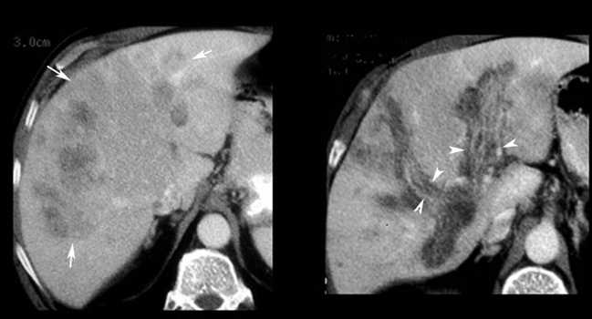
CT scan in a patient with Multicentric hepatoma
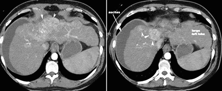
CT scan in another patient with Hepatoma
Arrowheads point to the enhancing mass. Note the lobulated margins of the liver, lower density than spleen and ascites indicating underlying cirrhosis.
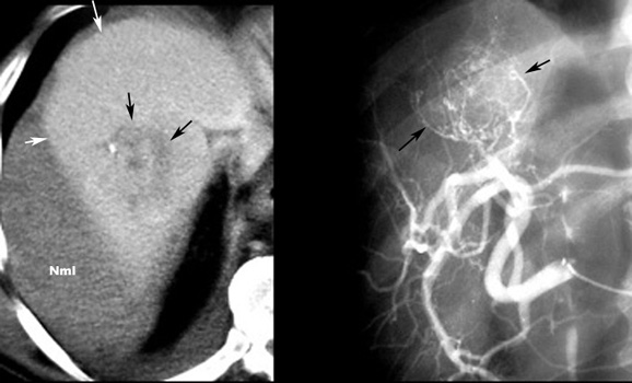
Hepatoma with hemorrhage
- NML is the normal liver density. White arrows point to increased density of the liver from hemorrhage (blood appears white on CT).
- Black arrows point to the hepatoma. Note tiny calcification in the tumor.
- Black arrows in the angiogram show the hypervascular tumor.
|
|
Hepatoma
- Arrows: Tumor
- Arrowheads: Tumor extends to portal vein
- Portal veins dilated with intraluminal tumor. Portal veins in liver appears dark on CT because it does not enhance with contrast.
MR Imaging
Single, multiple masses or diffuse involvement
- T1 weighted images
- Heterogeneous, isointense to hyperintense
- Hemorrhage
- Fat
- Necrosis
- Calcification
- T2 weighted images
- Most hyperintense, may be isointense
- Vascular invasion
- Enhancement reflects vascularity and necrosis
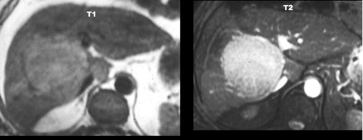
Hepatoma
MR shows a mass that has low signal intensity on T1 and high signal on T2.
Liver Cyst
Computed Tomography
- Oval, well defined
- Imperceptible or thin wall
- Water density
- No enhancement
- Wall calcification
- Septa
Sonography
- Well defined, anechoic
- May be echogenic due to fluid content
Magnetic Resonance
- T1 hypointense T2 marked hyperintense
- May be indistinguishable from hemangioma without IV contrast
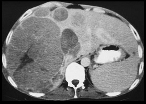
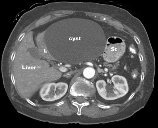
Liver Cyst
CT with IV contrast
Large cyst right lobe of liver.
- Oval, well defined
- Imperceptible or thin wall
- Water density
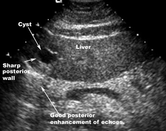
Liver Cyst
Ultrasound
Findings:
- An echoic mass: Cyst
- Sharp posterior wall
- Good posterior enhancement of echoes
- Ultrasound features that are specific for a cyst include an echolucent mass with a well-defined thin wall and increased through-transmission.
- Lesions which show these features need no further evaluation.
- Small (< 1.0 cm) cysts may be difficult to characterize with confidence.
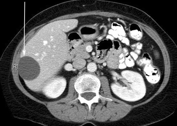
What are the imaging findings of liver metastases?
- Single lesion
- Multiple hypodense lesions
- Hypervascular lesions
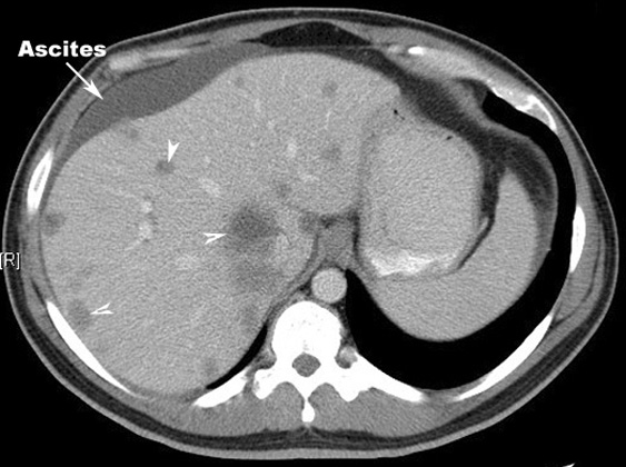
Liver metastasis
Multiple hypodense lesions seen in the liver with no significant contrast enhancement.
Primary: Colon carcinoma
Discuss the utility of imaging procedures for detection of liver metastases.
CT
- CT scan is the imaging procedure of choice to evaluate liver for metastases.
- Hypervascular metastases may be difficult to detect on CT scans performed with a single phase technique. Triphasic scans should be done.
- CT arterial portography can improve sensitivity for metastatic lesions, albeit with lower specificity.
Ultrasound
- Is most useful intraoperatively, during which the transducer is placed directly upon the liver surface to recognize liver metastases.
- Transabdominal ultrasonography is inferior in sensitivity for liver masses to CT or MRI.