Bowel perforation / Pneumoperitoneum
Findings:
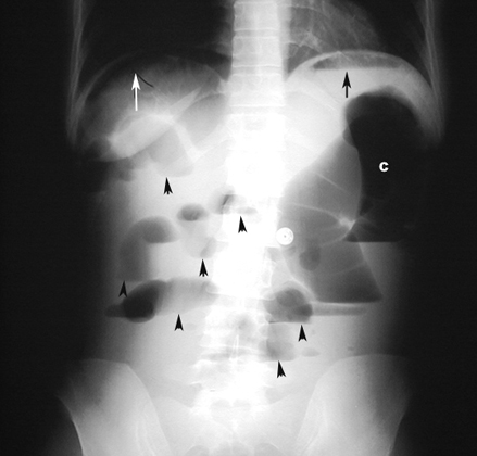
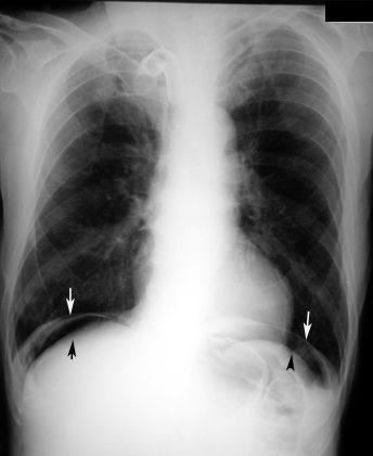
- Upright film of abdomen demonstrates air under the right hemidiaphragm (white arrow).
- Arrowheads point to multiple bowel loops with air fluid levels.
- Black arrow points to air fluid level in stomach.
|
Bowel perforation / PneumoperitoneumFindings:
|
 |
Bowel perforation / PneumoperitoneumFindings:
|
|
|
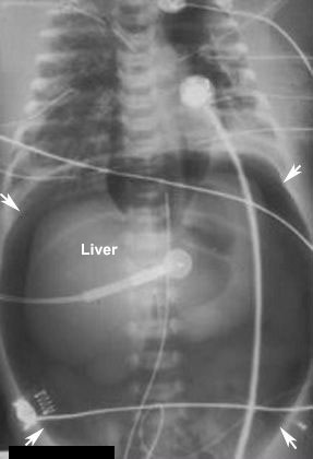
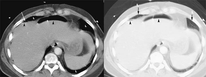
Large PneumoperitoneumFindings:
|
|
|
PneumoperitoneumFindings:
|
 |
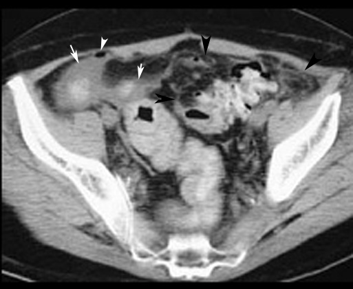
Bowel perforation / Pneumoperitoneum |
|
|
|
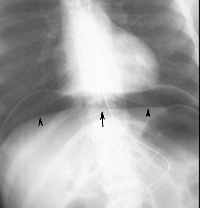
Pneumoperitoneum
|