|
UNDER CONSTRUCTION |
|
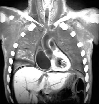
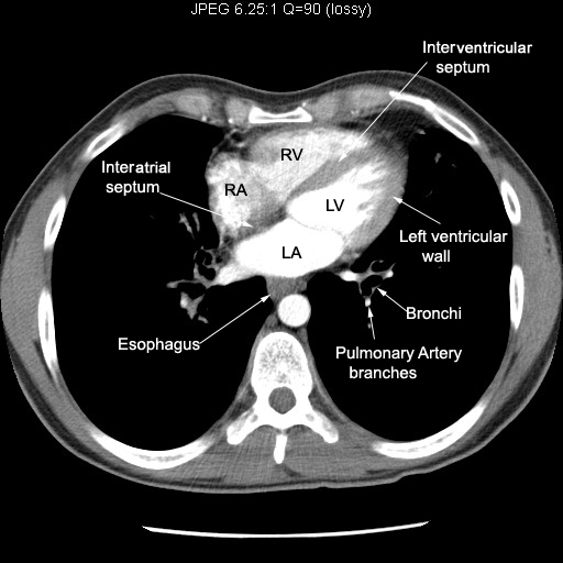
| Heart | |
| Right atrium | |
| Block Section | Identify right atrium |
| CT1 | Follow SVC entering right atrium. |
| CT2 | Note the relationship of right atrium to other chambers of heart. |
| CT3 | Note which heart border, the right atrium forms |
| CT4 | Right atrium emptying into right ventricle. |
| CXR | Identify the location of right atrium |
| Angiogram | |
| Left atrium | |
| Pulmonary veins entering left atrium | |
| CT 3 | Left atrium emptying into left ventricle |
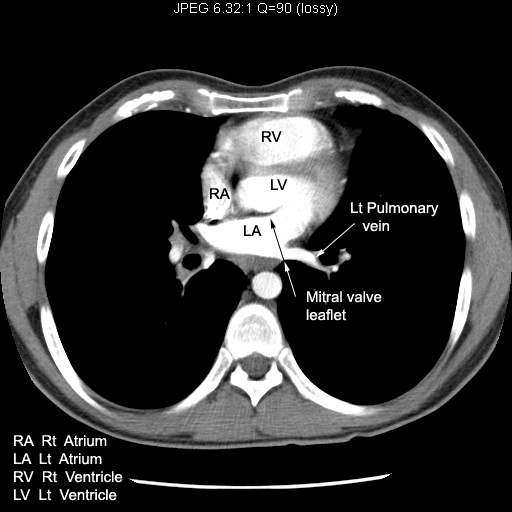
| CT 2 | Note mitral valve leaflet. Note its location. |
| CT4 | Relationship of LA to Esophagus |
| Angiogram | |
| Right ventricle | |
| Block Section | |
| Block Section | |
| CT 1 |
Note the relationship of right ventricle to Sternum. |
| CT 2 | Note the relationship to other chambers of heart |
| CT 3 | Right atrium emptying into right ventricle. |
| CT 4 | Note the interventricular septum |
| CXR | Locate right ventricle |
| Angiogram | |
| Left ventricle | |
| CT1 |
Note left ventricular wall thickness and Inter ventricular septum |
| CT 2 | Forms left heart border |
| CT3 | Origin of Aorta |
| CXR | Locate left ventricle |
| Angiogram | |
| Aorta | |
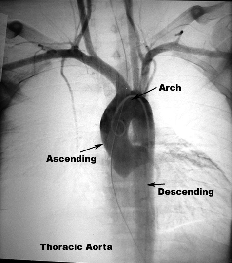
| Thoracic Aorta | |
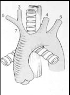
| Drawing | Follow Aorta from Aortic valve, ascending, arch and descending portions of Aorta. |
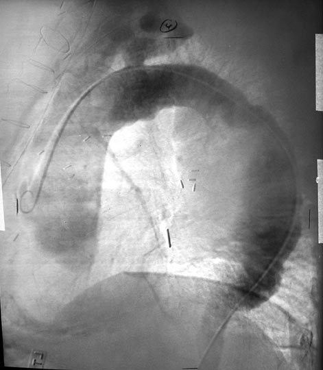
| Angiogram | Follow Aorta from Aortic valve, ascending, arch and descending portions of Aorta |
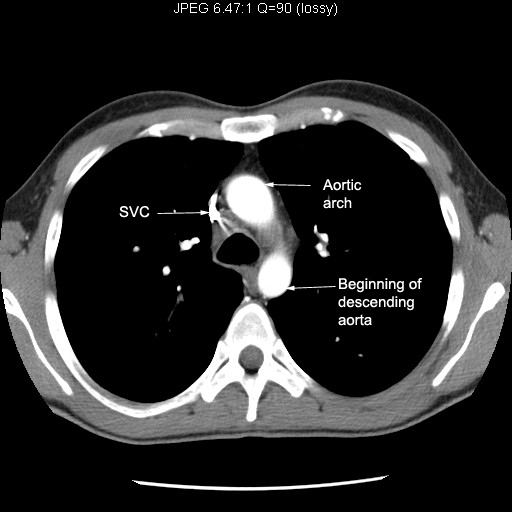
| CXR | Various portions of Aorta |
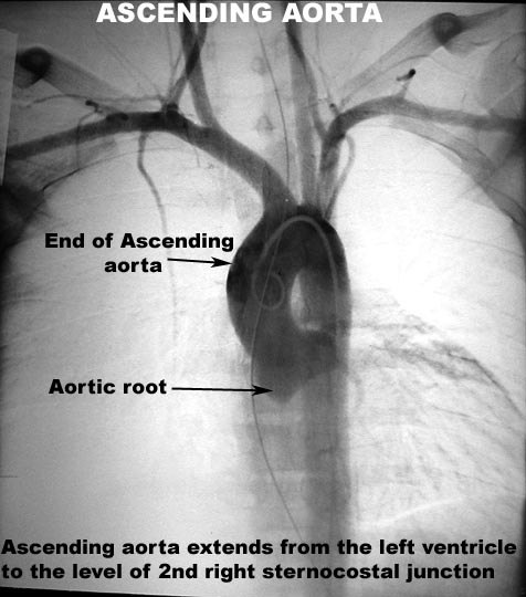
| Ascending aorta | |
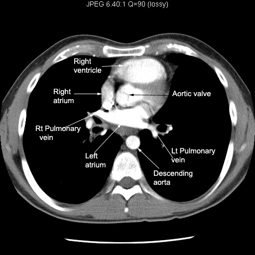
| CT 5 | Aortic valve |
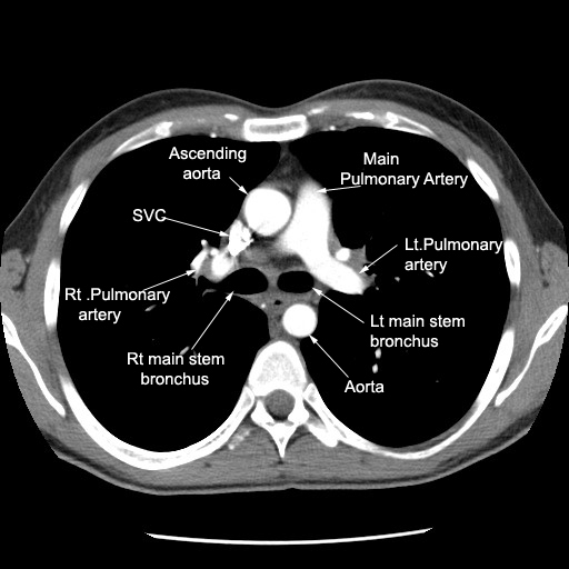
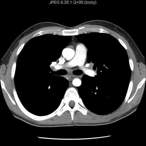
| CT 4 | Main pulmonary artery is to left of aortic root. |
| Angiogram | Ascending Aorta Start and end |
| Angiogram | Branches of Ascending Aorta |
| CXR | Location of ascending Aorta |
| Lateral | Location of ascending Aorta |
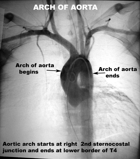
| Arch of aorta | |
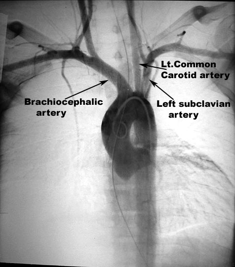
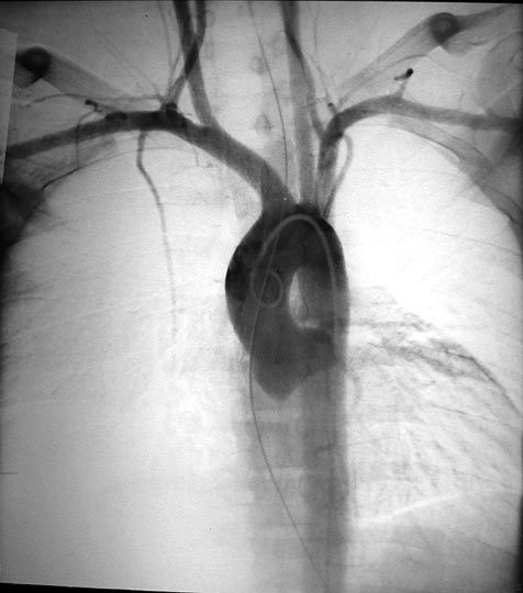
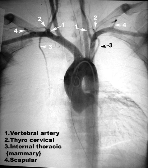
| Image1 | Major branches of Thoracic Aorta. Follow the major branches of Aortic arch to neck. |
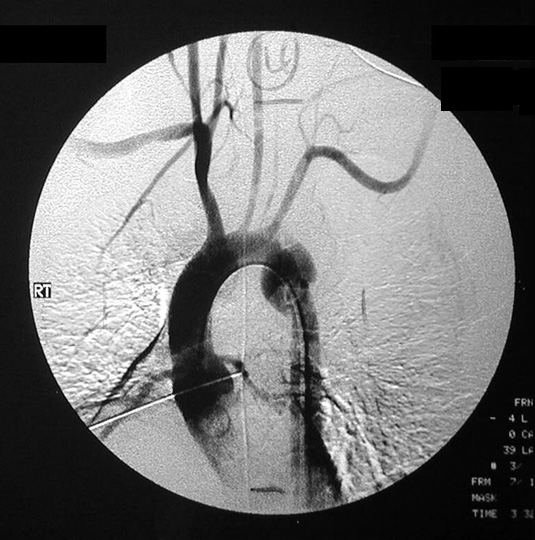
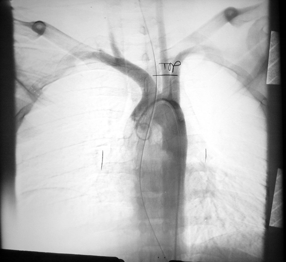
| Angiogram1 | Aorta and brnaches |
| Angiogram | Aorta and branches |
| Image4 | Aorta and branches |
| Image2 | Branches |
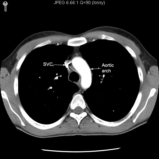
| CT 3 | Start of aortic arch |
| CT 2 | Aorta arching across mediastinum |
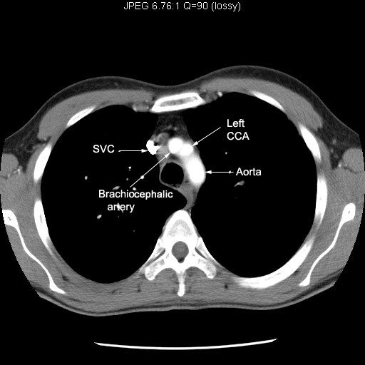
| CT 1 | Major branches on the surface of aortic arch |
| Lateral chest | Locate Aortic arch |
| CXR | Locate Aortic arch |
| Descending aorta | |
| Angiogram1 | Aorta and branches |
| Image2 | Aorta and branches |
| CT 6 | Relation to Esophagus, left pleura and Vertebra |
| CT 7 | Relation to Esophagus, left pleura and Vertebra |
| CT 8 | Relation to Esophagus, left pleura and Vertebra |
| CXR | Locate descending Aorta |
| Lateral | Locate descending Aorta |
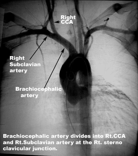
| Brachio cephalic artery | |
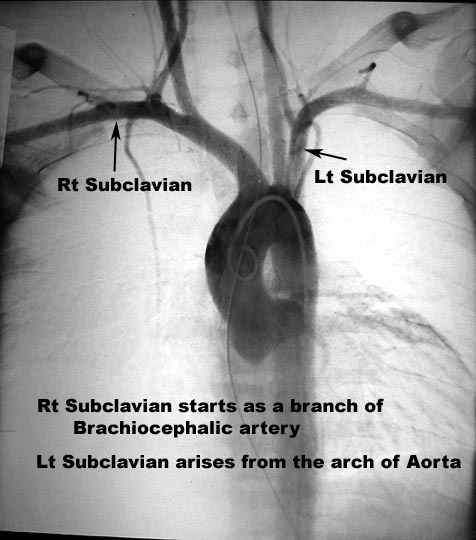
| Angiogram | Aorta and branches |
| CT 1 | Starting from the arch of aorta |
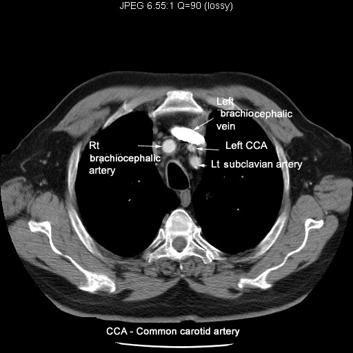
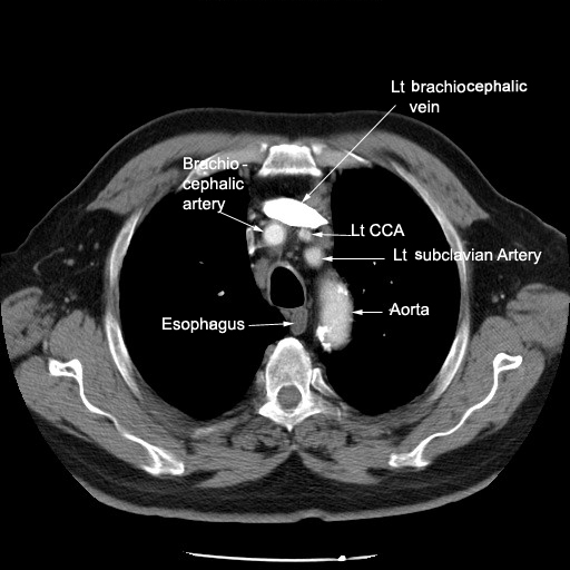
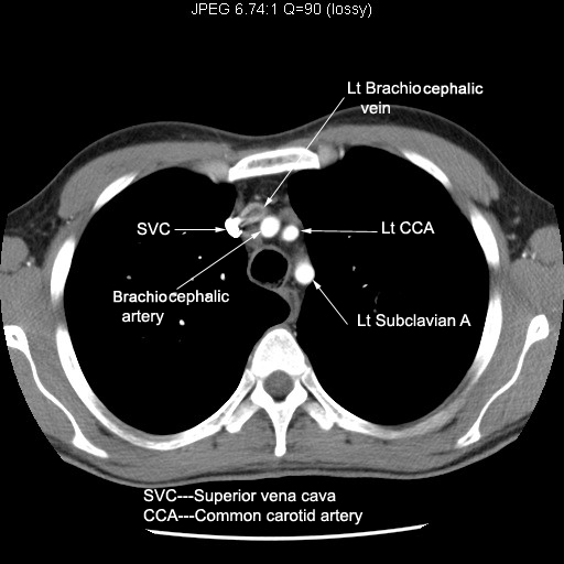
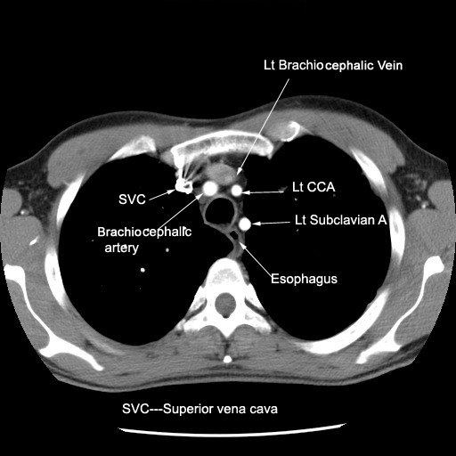
| CT3 | Relation to brachiocephalic vein |
| CT4 | Relation to brachiocephalic vein |
| CT 2 | Relation to brachiocephalic vein |
| CT 3 | Relation to brachiocephalic vein |
| CT 4 | Divides into right common carotid and right subclavian arteries |
| CT2 | . |
| Left Common carotid artery | |
| Angiogram1 | Aorta and brnaches |
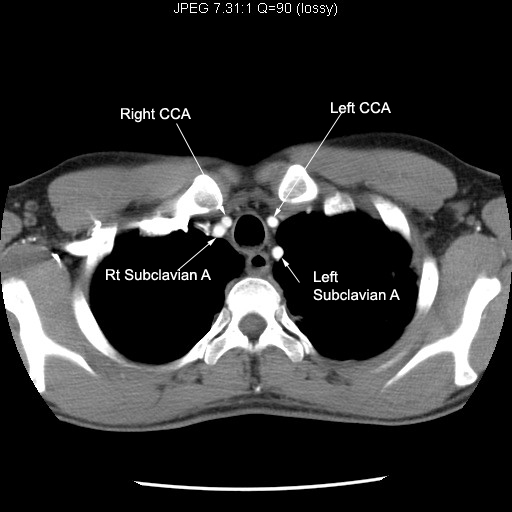
| CT 1 | Starting from the arch of aorta |
| CT 2 | Middle branch of the arch of aorta |
| CT 3 | Middle branch of the arch of aorta |
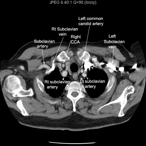
| CT 4 | Courses towards neck |
| CT2 | Courses into neck |
| CT3 | Relation to left brachiocephalic vein |
| CT4 | Relation to left brachiocephalic vein |
| Angiogram | Origin |
| Angiogram | Brnaches |
| CT 2 | Lat branch off of arch of aorta |
| CT 3 | Relation to trachea and esophagus |
| CT 4 | Relation to trachea and esophagus |
| CT2 | Coursing into neck |
| CT3 | |
| CT4 | |
| CT 5 | Coursing into neck |
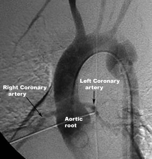
| Coronary artery | |
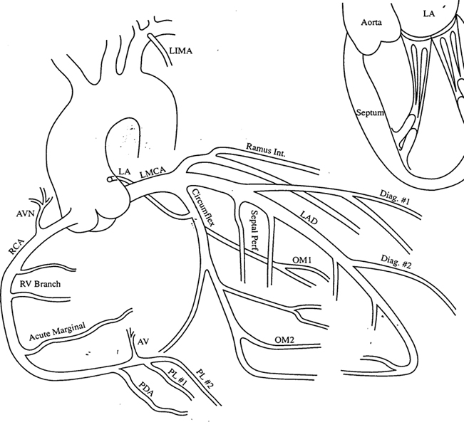
| Drawing | Coronary artery and its branches |
| Angiogram | Branches of Ascending Aorta |
| Angiogram | Left Coronary artery |
| Angiogram | Right Coronary artery |
| Angiogram | Branches of LCA |
| Angiogram | Branches of RCA |
| Superior Vena cava | |
| CT1 | Left joining right brachiocephalic vein to form SVC |
| CT2 | Left joining right brachiocephalic vein to form SVC |
| CT3 | SVC |
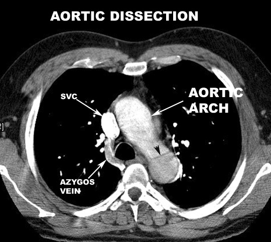
| CT4 | Relationship to Aorta and right pulmonary artery |
| CT5 | SVC drained into right atrium |
| Hema41a | Azygous vein joining SVC |
| Angiogram | |
| CXR | Locate SVC |
| Brachiocephalic veins | |
| CT1 | Internal jugular vein joins subclavian vein to form brachiocephalic vein |
| CT2 | Internal jugular vein joins subclavian vein to form brachiocephalic vein |
| CT3 | Left brachiocephalic crosses mediastinum towards right brachiocephalic vein |
| CT4 | Right brachiocephalic descends vertically |
| CT5 | Left joins right brachiocephalic vein to form SVC |
| CT6 | Left joins right brachiocephalic vein to form SVC |
| CT7 | Left joins right brachiocephalic vein to form SVC |
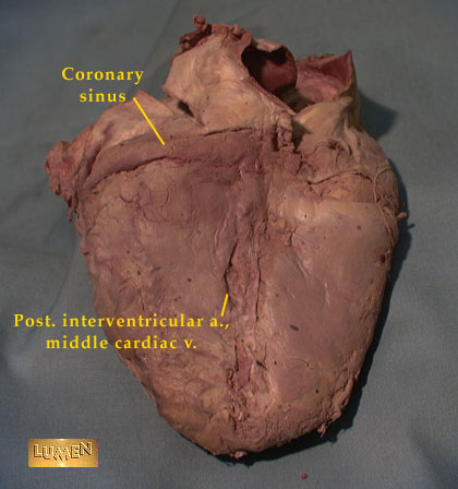
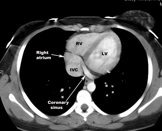
| Heart | Coronary sinus |
| Sandy2 | Coronary sinus in the atrioventricular groove and entering right atrium |
| . | . |
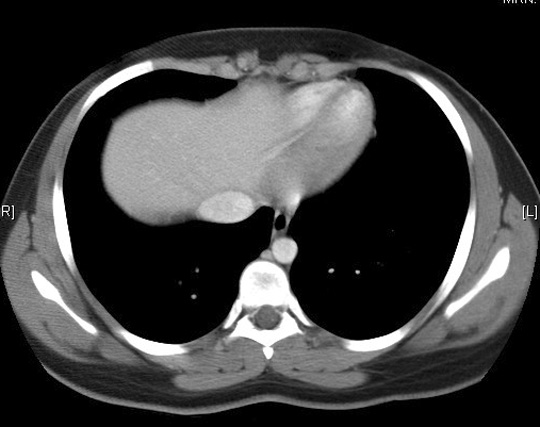
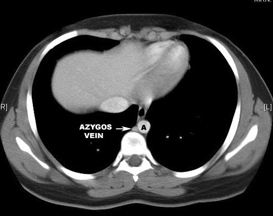
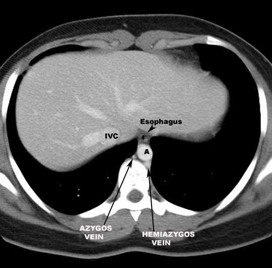
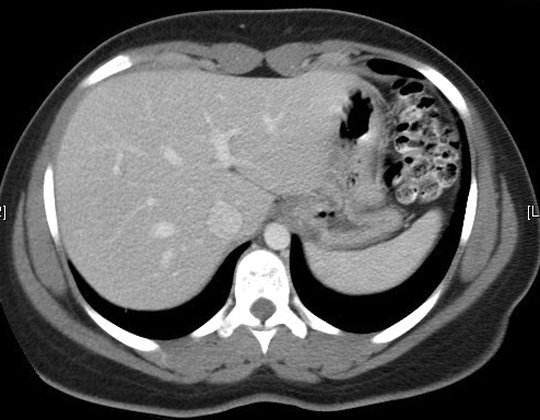
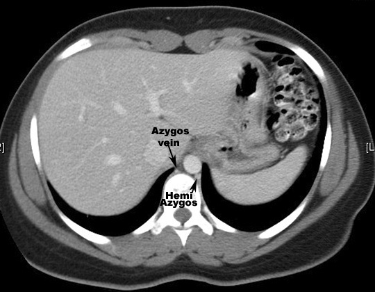
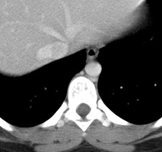
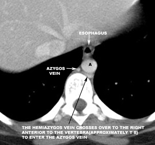
| gi/Sandy1 | Azygous vein relation to Aorta, Vertebra and Esophagus |
| Sandy3 | Azygous vein relation to Aorta, Vertebra and Esophagus |
| Sandy6 | Azygous vein relation to Aorta, Vertebra and Esophagus |
| Sandy7 | Azygous vein relation to Aorta, Vertebra and Esophagus |
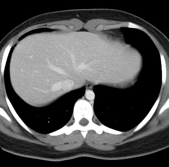
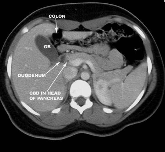
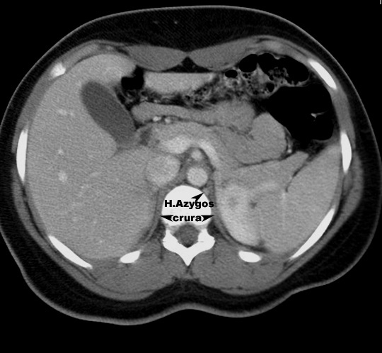
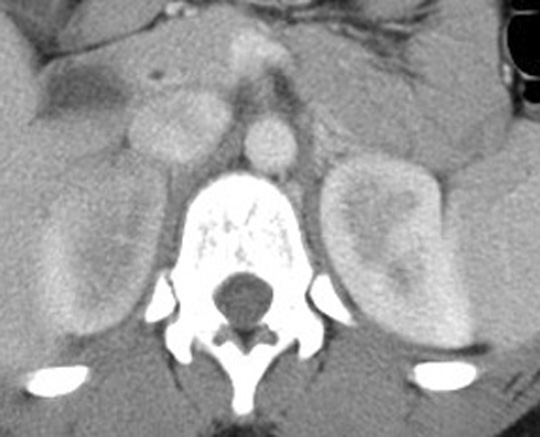
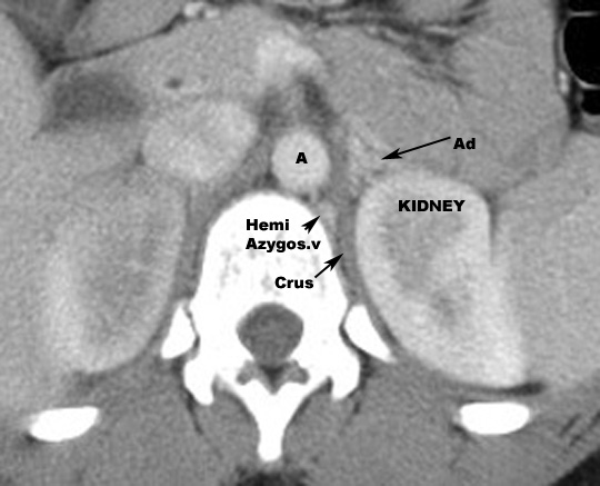
| Sandy12b | Hemiazygous vein relation to Aorta, Vertebra and diaphragmatic crus |
| Sandy14b | Hemiazygous vein relation to Aorta, Vertebra and diaphragmatic crus |
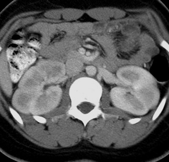
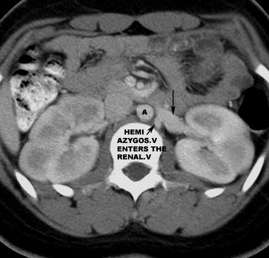
| Sandy19b | Hemiazygous connecting to left renal vein in this case. |
| Sandy5b | Hemiazygous crosses over to join Azygous vein |
| Hema41a | Azygous joining SVC |
| CXR | Locate Azygous vein |
| Inferior vena cava | |
| CXR | Locate IVC |
| Lateral | Locate IVC |
| CT | IVC entering RA |
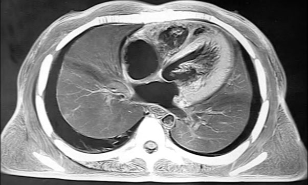
| Pulmonary artery | |
| Block Section | |
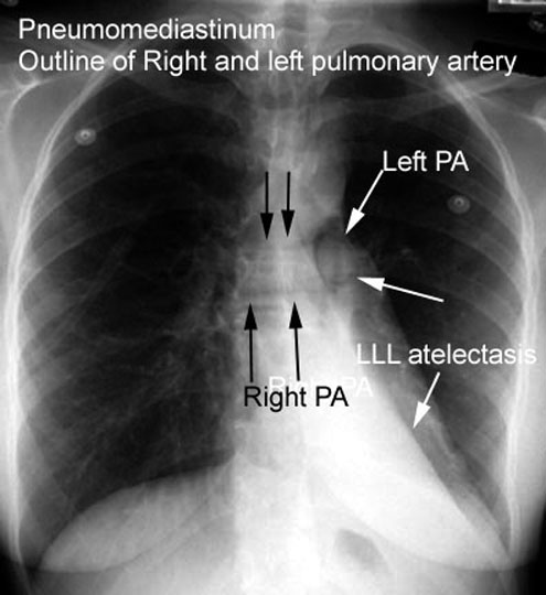
| CXR | |
| CXR | |
| CT 1 | To left of Aorta |
| CT 2 | Divides into left and right pulmonary arteries |
| CT 3 |
Pulmonary artery branches in the lungs. |
| Angiogram | Main Pulmonary artery |
| Angiogram | Right Pulmonary artery |
| Angiogram | Left Pulmonary artery |
| Angiogram | Lateral view |
| CXR | Right and left |
| Lateral | Locate left and right pulmonary arteries |
| Pulmonary veins | |
| CT 1 | Pulmonary veins entering left atrium. |
| CT 2 |
Left Pulmonary veins entering left atrium. |
| CXR | Locate pulmonary veins |