
|
Granulomatous Disease |

|
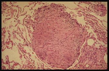
This noncaseating granuloma in the lung is the characteristic lesion of sarcoidosis but is not diagnostic of the disease. Noncaseous granulomas have a wide variety of causes other than sarcoidosis. |
There are many and varied causes of granulomatous disease. All of these etiologies, however, produce a similar distinctive histologic pattern called chronic granulomatous inflammation. Granulomas are small, 0.5 - 2mm collections of modified macrophages called epithelioid cells. These collections are usually encircled by lymphocytes and often contain giant cells.
Granulomas can be divided into the foreign body type and the hypersensitivity type. Hypersensitivity granulomas are the result of a cellular immunologic response to an antigen. In the case of sarcoidosis, the antigen or antigens provoking the response remain unknown.
Sarcoidosis is characterized by noncaseating granulomas. These are different than the caseating granulomas produced by other diseases, especially tuberculosis. Caseous necrosis is destruction of cells which are converted to amorphous greyish debris located centrally in granulomas. The term caseous ( L. caseus, cheese) refers to the gross appearance of caseous necrosis which resembles clumped, friable cheese.
Sites of disease in sarcoidosis are characterized by abnormal accumulation of helper T lymphocytes followed by granuloma formation. Symptoms and signs of the disease are due to the bulk of the granulomas altering organs and tissues. In most patients the granulomas resolve with no permanent sequelae. In chronic cases, however, inflammation can eventually lead to fibrosis and permanent organ dysfunction.


| Terrence Demos, M.D. |
Last Updated: March 14, 1996 Created: March 1, 1996 |