

|
Extrathoracic |
The reticuloendothelial mononuclear phagocyte system includes the bone marrow, lymph nodes, the liver, and the spleen. All of these sites are often involved by sarcoid granulomas. Spleen involvement producing hypersplenism and liver involvement producing cholestasis, cirrhosis, portal hypertension, Budd-Chiari syndrome, and liver failure have been reported, but are rare. Most patients with evident liver involvement have only biochemical abnormalities and hepatomegaly. .
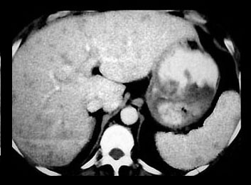
Hepatomegaly and splenomegaly occur in about one-third of patients. Liver lesions have been found in about 5% and spleen lesions 2-3 times as often in patients with sarcoidosis undergoing computed tomography examinations. The liver and spleen lesions most often appear as small widespread foci of decreased attenuation best seen when intravenous contrast material is utilized.
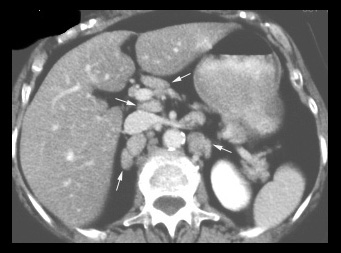
Subdiaphragmatic lymphadenopathy, both retroperitoneal and intraperitoneal, has been reported in up to one-half of these patients. The lymph nodes are usually discrete and mildly enlarged, but marked lymphadenopathy and large conglomerate nodes do occur.
| ABDOMINAL LYMPHADENOPATHY | SPLEEN & LIVER GRANULOMAS |
|---|---|
|
|
There are multiple enlarged paraaortic, paracaval, and porta hepatis lymph nodes (arrows). |
The small low attenuation lesions in the liver and spleen in sarcoidosis. |
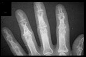
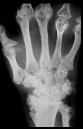
Acute polyarthritis is a common presenting symptom of sarcoidosis, but these patients do not have radiologic abnormalities. Visible bone lesions are associated with chronic disease and are rare unless the patient has skin lesions. The hands and feet are sites of lesions much more often than any other bone. The distal and middle phalanges are most often involved. The lesions are often few and inconspicuous, but rarely chronic disease leads to more severe and even destructive disease. Destructive granulomatous lesions can also occur in the facial bones and involve adjacent structures such as the paranasal sinuses, lacrimal glands, and optic nerve.
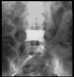
Next in frequency, but lagging far behind the hands and feet, is involvement of the skull and facial bones, especially the nasal bone; vertebrae; ribs; the pelvis and the ends of long bones. Most of the lesions in these sites are osteolytic, but sclerotic lesions do occur in the ribs, pelvis, and spine.
| PUNCHED OUT LYTIC LESIONS | LACY TRABECULAR PATTERN | DEFORMING LESIONS |
|---|---|---|
|
|
|
Focal osteolytic lesions in the fingers are the most common abnormality. The lesions in this patient are larger than usual. |
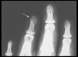
Osteolysis has left a lacy trabecular pattern in this phalanx (arrow). |
Advanced sarcoidosis with numerous osteolytic lesions of the distal forearm, wrist, and bones of the hand cause gross deformity. |
| SCLEROTIC LESION | SCLEROTIC LESIONS, NONSPECIFIC | NASAL BONE LESION |
|---|---|---|
|

|
|
Sclerotic sarcoid lesions are rare and are most often in the axial skeleton. |
Focal sclerosis (arrows) of distal phalanges is an unusual and nonspecific manifestation of sarcoidosis. |
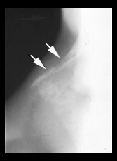
Nasal sarcoidosis can lead to osteolysis of the nasal bone (arrows). |
Aside from the salivary glands, gastrointestinal lesions are rare. Esophageal lesions are extremely rare.
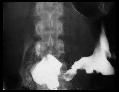
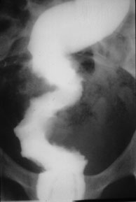
Antral narrowing is the most common type of stomach lesion. This narrowing is indistinguishable from other more common causes of narrowing including carcinoma and other granulomatous diseases such as Crohn's disease, tuberculosis, syphilis, and fungal disease. Small bowel sarcoidosis can resemble Crohn's disease or malignancy as can colon lesions.
| GASTRIC SARCOID | COLONIC SARCOID |
|---|---|
|
|
The granulomatous process involves the gastric antrum leading to irregular nonspecific narrowing. |
Irregular narrowing of the rectosigmoid due to sarcoidosis has the appearance of inflammatory disease or malignancy . |
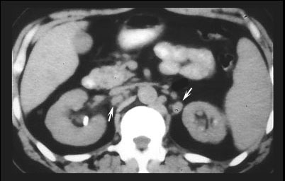
Findings in the kidney are related to hypercalcemia and hypercalciuria due to sarcoidosis. A very small number of patients who have chronic calcium elevation develop kidney stones. Rarely calcium is deposited in the renal tissue and collecting tubules producing the characteristic multiple focal medullary calcifications of nephrocalcinosis. This pattern is indistinguishable from nephrocalcinosis due to other causes, but most patients with sarcoidosis have clearly identifiable involvement of other organ systems.
| NEPHROCALCINOSIS |
|---|
|
There are multiple calcifications of the kidneys (nephrocalcinosis) as a result of hypercalcemia. Note enlarged retroperitoneal lymph nodes (arrows) which is a common finding in sarcoidosis. |

| Terrence C. Demos, M.D. |
Last Updated: March 14, 1996 Created: March 1, 1996 |