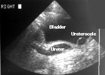
A four-year-old girl has had two urine infections. Her physician ordered the ultrasound seen on the right. Hydronephrosis was seen in the right kidney (image not shown). This is a longitudinal view of the distal right ureter and bladder. The left side of the image (as you face it) is cephalad; the right side is caudal. Notice that the distal ureter is enlarged. Just to the right of the ureter (caudal) a cystic structure lies within the bladder. This is the dilated portion of the distal ureter within the bladder, a ureterocele. The ureter and ureterocele, in fact, are not separated. The ureteric lumen is widened all the way into the bladder, but it is somewhat tortuous so that the ultrasound image, seeing only one plane, seems to show them separated.
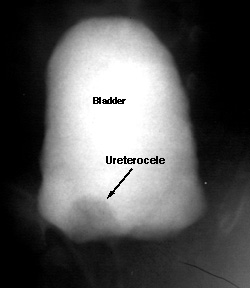
A voiding cystourethrogram (shown at left) shows a filling defect (darker gray area) at the right bladder base. This is the ureterocele seen on the ultrasound. Notice that there is no reflux. Some ureteroceles do reflux, allowing urine to move retrograde from the bladder up the ureter.
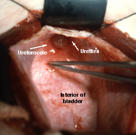
This ureterocele was removed surgically by opening the bladder and reconnecting the ureter with the bladder (a ureteroneocystostomy). This surgical photo was taken from the patient's chest looking caudad into the base of the opened bladder.