
|
|
Lateral Chest Radiograph
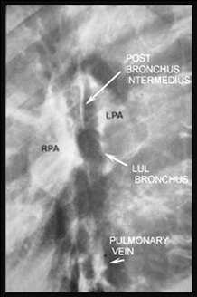
Five normal hilar structures are usually visible.
| LABELED LATERAL VIEW OF NORMAL HILUS | UNLABELED NORMAL HILUS |
|---|---|
|
|
Five labeled normal structures. |
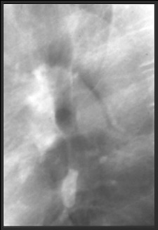
Normal structures in another patient. |
The small oval pulmonary vein is the only normal round density inferior to the hila. If there a large infrahilar increased density, this is lymphadenopathy or a lung mass. If there is a "doughnut" of increase density surrounding the "hole" which is the end on left upper lobe bronchus, this indicates more advanced hilar lymphadenopathy.
| LATERAL VIEWS | OF HILAR | LYMPHADENOPATHY |
|
|
|
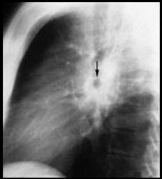
This patient has hilar lymphadenopathy seen as a "donut" of soft tissue density surrounding the "doughnut hole" which is the end on left upper lobe bronchus (arrow). |
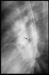
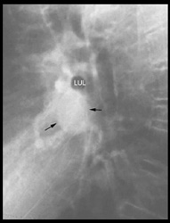
A lopsided soft tissue "doughnut" surrounds the end on LUL bronchus (arrow). This is more advanced hilar lymphadenopathy. (The opaque line is a nasogastric tube.) |
Here there is an area of infrahilar soft tissue density that is much too large to be a normal pulmonary vein. This is less advanced hilar lymphadenopathy.(LUL=left upper lobe bronchus) |


| Terrence C. Demos, M.D. |
Last Updated: March 14, 1996 Created: March 1, 1996 |